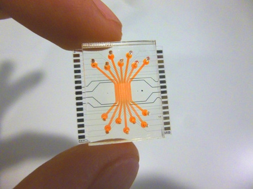
Microfluidic device that reproduces the blood-retinal barrier
The use of In vitro testing with living cells as an alternative to animal research has limitations like the difficulty to reproduce the interaction of cells. To overcome it, scientists are working on the development of systems that simulate and reproduce functions of tissues and organs in conditions very similar to reality. They are called organ-on-a-chip, which include microenvironments and microarchitectures that simulate the state of tissues and living organs.
Scientists of NANBIOSIS Unit 8 have published in an article, cover of the magazine “Lab on a Chip”, the “proof of concept” of a microfluidic device that reproduces the blood-retinal barrier, that is, a microchip that allows us to reproduce what happens ” in vivo ‘in the retina. This device can be an essential tool that revolutionizes experimentation ‘in vitro’.
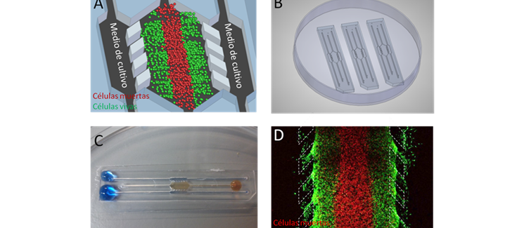
José Yeste, researcher of the CIBER-BBN, explains that the micro device consists of several parallel compartments, in which different types of cells have been cultivated to emulate the structure of cellular layers of the retina. They are endothelial cells, that is, they form the internal part of the barrier, in contact with the blood capillaries, through which oxygen and nutrients reach the retina. In addition, it is also composed of neuronal cells (which form the neuroretina), and pigment epithelial cells, which constitute the outer layer. The compartments are interconnected in their lower part by a network of micro-grooves, so as to allow an intercellular communication through the exchange of signalling molecules between cells. Thus, cells can send their signals to others and interact, much like they would in a living organism. In addition, the micro device allows the endothelial cells to be subjected to the mechanical stimulus induced by the flow to emulate a more physiological microenvironment.
“Within the body, the endothelial cells that line the inside of blood vessels are subject to the mechanical stimulation of blood circulation. In cell cultures that do not reproduce this flow, the cells are as ‘lethargic’, and do not respond in the same way they would in real conditions, “explains Rosa Villa, Scientific Director of NANBIOSIS Unit 8 and leader of the group of Biomedical Applications of the Microelectronics Institute of Barcelona of the CSIC.
Scientists have evaluated the correct formation of the blood-retinal barrier by performing permeability, electrical resistance tests, as well as protein expression of tight junctions between cells. These tests were intended to verify that the barrier is well formed, that it has closed but maintains the natural permeability, sufficient to allow the passage of nutrients and oxygen, and that the cells are in contact and interact with each other.
This work has been developed in the ICTS NANBIOSIS, more specifically in Unit 8 of Micro-Nano Technology located in the IMB-CNM. It is also part of the results of the CIBER intramural project called Micro BRB: Microfluidic model of retinal neurovascular unit to identify new therapeutic targets in diabetic retinopathy (2016-2017) in wich also participates Unit 3 of NANBIOSIS
Article of reference:
A compartmentalized microfluidic chip with crisscross microgrooves and electrophysiological electrodes for modeling the blood–retinal barrier. Jose Yeste, Marta arcía-Ramírez, Xavi Illa, Anton Guimerà, Cristina Hernández, Rafael Simó and Rosa Villa. DOI: 10.1039/C7LC00795GLab Chip, 2018, 18, 95-105


