+34 620 10 75 37info@nanbiosis.com

Singular

Singular

U19-S03. Clinical Analysis Laboratory

Clinical Analysis Laboratory

The clinical analysis laboratory that performs a wide variety of analysis: (biochemistry, hematology, coagulation, urine analysis, blood gas analysis, different biomarkers, hormones, allergens) in various animal species (rat, mouse, rabbit, pig, sheep, dog, and cat). Specific methodologies are used in each animal species, having all techniques validated according to their physiological and pathological identity. It also has a formulation laboratory equipped to formulate, reconstitute and guarantee the correct storage of the test products used in these studies.

Customer benefits

The laboratories are certified with ISO-9001 and Good Laboratory Practices (GLP), strict quality standards that allow the production of high-precision results.
Also, the studies can be performed under regulatory requirements too, since the performing institution is Certified for Good Laboratory Practices.
Therefore, studies can be carried out in compliance with the strict guidelines of regulatory agencies, ensuring the reliability and traceability of all results and tests carried out in their different services.

Target customer

The services offered in this unit may be of interest to different companies and laboratories that work within the pharmaceutical industry. Companies whose objective is to test possible candidates for molecules, drugs or medical devices.
With this service, companies and laboratories ensure that they report reliable and reproducible results in all their studies, guaranteeing the traceability and viability of analyzes obtained.

References

  1. J Bote, et al. Novel ex-vivo database of a murine model of colorectal cancer using optical coherence tomography. Surg Endosc (2022) 36:S325–S674
  2. V Lucas-Cava, et al. Prostatic artery occlusion: Initial findings on pathophysiological response in a canine prostate model. Translational Andrology and Urology. Transl Androl Urol 2022.
  3. Soria F, et al. Heparin coating in biodegradable ureteral stents does not decrease bacterial colonization-assessment in ureteral stricture endourological treatment in animal model. Transl Androl Urol. 2021 Apr;10(4):1700-1710.
  4. Baez-Díaz C, et al. Microencapsulated Insulin-Like Growth Factor-1 therapy improves cardiac function and reduces fibrosis in a porcine acute myocardial infarction model. Sci Rep. 2020 Apr 28;10(1):7166.
  5. Moreno-Lobato, B, et al. Use of nanomedicine in preclinical wound healing studies. FarmaJournal; Salamanca T 4, N.º 1, (Feb 2019): 178.
  6. Picado Román, N, et al. Drugs most used in experimental animals in a Research Center. FarmaJournal; Salamanca T 4, N.º 1, (Feb 2019): 236.
  7. Moreno-Lobato B, et al. Analytical Validation Study of Hematological Parameters under Good Laboratory Practice Regulations in Different Laboratory Animal Species. Thromb Haemost Res. 2019; 3(1): 1018.
  8. Ballestín A, et al. A Pre-clinical Rat Model for the Study of Ischemia reperfusion Injury in Reconstructive Microsurgery. J Vis Exp. 2019 Nov 8;(153).
  9. R Blázquez , et al. Altered hematological, biochemical and immunological parameters in a porcine myocardial infarction model: predictive biomarkers for the severity of myocardial infarction. Veterinary Immunology and Immunopathology 205 (2018) 49–57.
  10. Vela FJ, et al. Evaluation of antigen-induced synovitis in a porcine model: Immunological, arthroscopic and kinetic studies. BMC Vet Res. 2017. Apr 7;13(1):93.
Read More

U14-S06. Lymphocyte proliferation assay

Lymphocyte proliferation assay

The lymphocyte proliferation assay service offers a robust platform for evaluating the proliferative responses of lymphocytes in vitro. Utilizing state-of-the-art stimulation protocols and specialized proliferation assays, this service enables precise quantification of lymphocyte activation and proliferation rates. By mimicking physiological conditions in a controlled laboratory setting, researchers can investigate the dynamic behavior of lymphocytes in response to various stimuli. This service plays a pivotal role in elucidating immune responses, assessing immune competence, and evaluating the efficacy of immunomodulatory interventions, ultimately contributing to advancements in both basic immunology and clinical therapeutics.

Customer benefits

U14 Cell Therapy Unit offers researchers precision, efficiency, and expertise, streamlining experiment. With stringent quality control, ISO 9001-2015 and GLP compliant, tailored solutions, and access to advanced equipment, researchers can focus on innovation and core objectives, driving scientific progress forward.

Target customer

This service is essential for organizations involved in Research and Development, including pharmaceutical companies, academic institutions for biomedical research, biotechnology firms for bioprocessing, and regulatory agencies for in vitro studies.

Additional information

Selected references:

  • de Pedro, Maria Ageles; Pulido, Maria; Álvarez, Veronica; Marinaro, Federica; Marchena, Ana Maria; Sanchez-Margallo, Francisco Miguel; Casado, Javier G.; Lopez, Esther.  2023.  Menstrual blood-derived stromal cells: insights into their secretome in acute hypoxia conditions. MOLECULAR MEDICINE. SPRINGER. 29-1. ISSN 1076-1551.  https://doi.org/10.1186/s10020-023-00646-1
  • Cristobal JI; Duque, FJ; Uson-Casaus J; Barrera R; López E; Perez-Merino EM.  2022.  Complete Blood Count-Derived Inflammatory Markers Changes in Dogs with Chronic Inflammatory Enteropathy Treated with Adipose-Derived Mesenchymal Stem Cells   Animals.  MDPI.  12-20.  ISSN 2076-2615.  https://doi.org/10.3390/ani12202798
  • Marinaro, Federica; Silva, Joana M; Barros, Alexandre A; et al; Lopez, Esther.  2021.  A Fibrin Coating Method of Polypropylene Meshes Enables the Adhesion of Menstrual Blood-Derived Mesenchymal Stromal Cells: A New Delivery Strategy for Stem Cell-Based Therapies.International journal of molecular sciences. MDPI. 22-24. ISSN 1422-0067. WOS (0) https://doi.org/10.3390/ijms222413385
  • de Pedro, Maria Angeles; Gomez-Serrano, Maria; Marinaro, Federica; et al; Lopez, Esther (AC); Casado, Javier G.(4/10).  2021.  IFN-Gamma and TNF-Alpha as a Priming Strategy to Enhance the Immunomodulatory Capacity of Secretomes from Menstrual Blood-Derived Stromal Cells   INTERNATIONAL JOURNAL OF MOLECULAR SCIENCES.  MDPI.  22-22.  ISSN 1422-0067.  WOS (0) https://doi.org/10.3390/ijms222212177
  • López E; Blazquez R; Marinaro F; et al; Casado JG. 2019. The Intrapericardial Delivery of Extracellular Vesicles from Cardiosphere-Derived Cells Stimulates M2 Polarization during the Acute Phase of Porcine Myocardial Infarction. STEM CELL REVIEWS AND REPORTS. SPRINGER. 16-3, pp.626-626. ISSN 2629-3269.  https://doi.org/10.1007/s12015-019-09926-y

Read More

U14-S05. Determination of cytokine concentration by flow cytometry

Determination of cytokine concentration by flow cytometry

The determination of cytokine concentration by flow cytometry service represents a cutting-edge approach to cytokine analysis within the realm of biomedical research and pharmaceutical investigation. Through the utilization of advanced flow cytometry techniques and specialized staining protocols, this service enables the precise quantification of intracellular cytokines across distinct cell subsets distinguished by surface markers. This service facilitates the elucidation of cytokine profiles within specific cell populations, shedding light on their roles in health, disease, and therapeutic interventions.

Customer benefits

U14 Cell Therapy Unit offers researchers precision, efficiency, and expertise, streamlining experiment. With stringent quality control, ISO 9001-2015 and GLP compliant, tailored solutions, and access to advanced equipment, researchers can focus on innovation and core objectives, driving scientific progress forward. By delving into the intracellular milieu, researchers gain invaluable insights into the intricate regulatory networks governing immune responses and cellular signaling pathways.

Target customer

This service is essential for organizations involved in Research and Development, including pharmaceutical companies, academic institutions for biomedical research, biotechnology firms for bioprocessing, and regulatory agencies for in vitro studies.

Additional information

Selected references:

  • de Pedro, Maria Ageles; Pulido, Maria; Álvarez, Veronica; Marinaro, Federica; Marchena, Ana Maria; Sanchez-Margallo, Francisco Miguel; Casado, Javier G.; Lopez, Esther.  2023.  Menstrual blood-derived stromal cells: insights into their secretome in acute hypoxia conditions. MOLECULAR MEDICINE. SPRINGER. 29-1. ISSN 1076-1551.  https://doi.org/10.1186/s10020-023-00646-1
  • Cristobal JI; Duque, FJ; Uson-Casaus J; Barrera R; López E; Perez-Merino EM.  2022.  Complete Blood Count-Derived Inflammatory Markers Changes in Dogs with Chronic Inflammatory Enteropathy Treated with Adipose-Derived Mesenchymal Stem Cells   Animals.  MDPI.  12-20.  ISSN 2076-2615.  https://doi.org/10.3390/ani12202798
  • Marinaro, Federica; Silva, Joana M; Barros, Alexandre A; et al; Lopez, Esther.  2021.  A Fibrin Coating Method of Polypropylene Meshes Enables the Adhesion of Menstrual Blood-Derived Mesenchymal Stromal Cells: A New Delivery Strategy for Stem Cell-Based Therapies.International journal of molecular sciences. MDPI. 22-24. ISSN 1422-0067. WOS (0) https://doi.org/10.3390/ijms222413385
  • de Pedro, Maria Angeles; Gomez-Serrano, Maria; Marinaro, Federica; et al; Lopez, Esther (AC); Casado, Javier G.(4/10).  2021.  IFN-Gamma and TNF-Alpha as a Priming Strategy to Enhance the Immunomodulatory Capacity of Secretomes from Menstrual Blood-Derived Stromal Cells   INTERNATIONAL JOURNAL OF MOLECULAR SCIENCES.  MDPI.  22-22.  ISSN 1422-0067.  WOS (0) https://doi.org/10.3390/ijms222212177
  • López E; Blazquez R; Marinaro F; et al; Casado JG. 2019. The Intrapericardial Delivery of Extracellular Vesicles from Cardiosphere-Derived Cells Stimulates M2 Polarization during the Acute Phase of Porcine Myocardial Infarction. STEM CELL REVIEWS AND REPORTS. SPRINGER. 16-3, pp.626-626. ISSN 2629-3269.  https://doi.org/10.1007/s12015-019-09926-y
Read More

U14-S04. Cytokine quantification assays

Cytokine quantification assays

The cytokine quantification assays service is at the forefront of cytokine analysis in biomedical research and pharmaceutical development. Leveraging advanced immunoassay techniques and specialized expertise, this service offers precise quantification of cytokines present in cell culture supernatants or animal fluids. Through meticulous analysis, researchers gain valuable insights into the intricate signaling networks and immune responses underlying various physiological and pathological processes. From characterizing inflammatory cascades to evaluating therapeutic interventions, our service provides a robust platform for investigating cytokine dynamics and their implications for health and disease.

Customer benefits

U14 Cell Therapy Unit offers researchers precision, efficiency, and expertise, streamlining experiment. With stringent quality control, ISO 9001-2015 and GLP compliant, tailored solutions, and access to advanced equipment, researchers can focus on innovation and core objectives, driving scientific progress forward.

Target customer

This service is essential for organizations involved in Research and Development, including pharmaceutical companies, academic institutions for biomedical research, biotechnology firms for bioprocessing, and regulatory agencies for in vitro studies.

Additional information

Selected references:

  • de Pedro, Maria Ageles; Pulido, Maria; Álvarez, Veronica; Marinaro, Federica; Marchena, Ana Maria; Sanchez-Margallo, Francisco Miguel; Casado, Javier G.; Lopez, Esther.  2023.  Menstrual blood-derived stromal cells: insights into their secretome in acute hypoxia conditions. MOLECULAR MEDICINE. SPRINGER. 29-1. ISSN 1076-1551.  https://doi.org/10.1186/s10020-023-00646-1
  • Cristobal JI; Duque, FJ; Uson-Casaus J; Barrera R; López E; Perez-Merino EM.  2022.  Complete Blood Count-Derived Inflammatory Markers Changes in Dogs with Chronic Inflammatory Enteropathy Treated with Adipose-Derived Mesenchymal Stem Cells   Animals.  MDPI.  12-20.  ISSN 2076-2615.  https://doi.org/10.3390/ani12202798
  • Marinaro, Federica; Silva, Joana M; Barros, Alexandre A; et al; Lopez, Esther.  2021.  A Fibrin Coating Method of Polypropylene Meshes Enables the Adhesion of Menstrual Blood-Derived Mesenchymal Stromal Cells: A New Delivery Strategy for Stem Cell-Based Therapies.International journal of molecular sciences. MDPI. 22-24. ISSN 1422-0067. WOS (0) https://doi.org/10.3390/ijms222413385
  • de Pedro, Maria Angeles; Gomez-Serrano, Maria; Marinaro, Federica; et al; Lopez, Esther (AC); Casado, Javier G.(4/10).  2021.  IFN-Gamma and TNF-Alpha as a Priming Strategy to Enhance the Immunomodulatory Capacity of Secretomes from Menstrual Blood-Derived Stromal Cells   INTERNATIONAL JOURNAL OF MOLECULAR SCIENCES.  MDPI.  22-22.  ISSN 1422-0067.  WOS (0) https://doi.org/10.3390/ijms222212177
  • López E; Blazquez R; Marinaro F; et al; Casado JG. 2019. The Intrapericardial Delivery of Extracellular Vesicles from Cardiosphere-Derived Cells Stimulates M2 Polarization during the Acute Phase of Porcine Myocardial Infarction. STEM CELL REVIEWS AND REPORTS. SPRINGER. 16-3, pp.626-626. ISSN 2629-3269.  https://doi.org/10.1007/s12015-019-09926-y
Read More

U11-S2. Formulation Lab

Read More

U20-S11. Histological porcessing, section and staining (Remote) OUTSTANDING

Read More

U20-S09. X-ray imaging in vitro and in vivo (Remote) OUTSTANDING

Read More

U13-S08. Atomic Force Microscopy indentation test

Atomic Force Microscopy indentation test

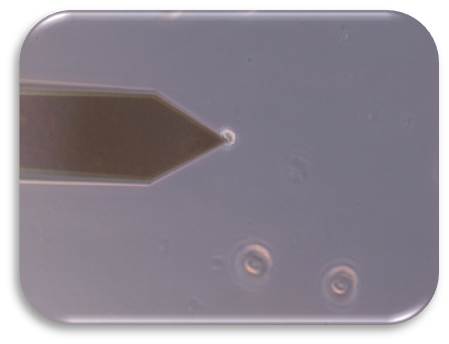
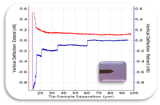
Atomic Force Microscopy can be used to measure the forces between the probe and the sample as a function of their mutual separation. This can be applied to perform force spectroscopy.

Customer benefits

Low forces on cells (nano-scale).

Target customer

Public and private research groups focused on mechanobiology

Additional information

Cantilever + cells
Force curve
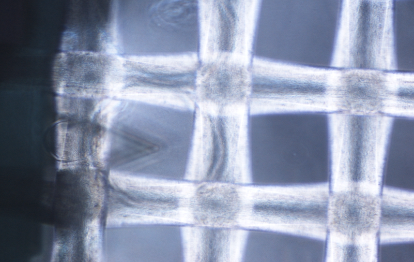
Capsules
AFM Software
Read More

U10-S9.

Characterization and development of pulmonar formulations

Encapsulation of therapeutic actives of interest in formulations for pulmonary administration with possibility of lyophilisation to obtain a powder. The service has two cutting-edge equipments for the characterization of pulmonary formulations:
SprayTec laser diffraction system: allows the measurement of spray particle and spray droplet size distributions in real-time for more efficient product development of sprays and aerosols, with robust and reproducible droplet size data.
Next Generation Impactor: has been designed specifically for the pharmaceutical industry for testing metered-dose inhalers, drypowder inhalers, nebulizers and nasal sprays. It consists of a high performance cascade impactor for classifying aerosol particles into micrometer size fractions, providing relevant information about their distribution in the respiratory tract.

Customer benefits

The formulations can be characterized, following SOPs, in terms of particle size, polydisperstity index and zeta potential. Importantly, this service can also offer real-time droplet size distribution and aerosol particles classification analysis by means of SprayTec and Next Generation Impactor (NGI) technology.

Target customer

  • Preclinical use for the characterization of the pulmonary formulation prior to its implementation in in vivo models.
  • Companies interested in characterizing their formulations or their pulmonary administration systems.

References

Moreno-Sastre M, Pastor M, Esquisabel A, Sans E, Viñas M, Fleischer A, Palomino E, Bachiller D, Pedraz JL. Pulmonary delivery of tobramycin-loaded nanostructured lipid carriers for Pseudomonas aeruginosa infections associated with cystic fibrosis. Int J Pharm. 2016 Feb 10;498(1-2):263-73. doi: 10.1016/j.ijpharm.2015.12.028.

Additional information

U10 Pulmonar formulations characterization.tif

Read More

U10-S8.

3D bio-impression of scaffolding for regenerative medicine

The principle of 3D bioprinting consists of selecting the most suitable biomaterials and cell types to prepare a Bioink that should be able to promote cell growth and differentiation and present appropriate mechanical properties of the target tissue.
This service possess a wide variety of 3D bioprinting techniques avaliable, such as extrusion, droplet, electrospining, electrowritting and stereolithography.

Customer benefits

One of the main characteristics of this additive manufacturing technique is its ability to bioprint the desired layers, with specific cell orientation , and desired morphology of the bioprinted 3D scaffold in order to ressemble, as much as possible, the tissue of interest. To achieve this goal, rheology, texturometry, printability and biological assays are carried out.

On the one hand, this technology can be employed to develop 3D scaffolds specific for the regeneration of particular tissues. On the other hand, this strategy offers a 3D environment that mimics the tissue/ organ of interest in order to test potential therapeutic tools, which goes in accordance with the implementation of the 3R principle (replace, reduce and refine).

Target customer

  • Preclinical use for in vitro and in vivo models.
  • Pharmaceutical industry (e.g. cosmetics)

References

  • Lafuente-Merchan M, Ruiz-Alonso S, García-Villén F, Zabala A, de Retana AMO, Gallego I, Saenz-Del-Burgo L, Pedraz JL. 3D Bioprinted Hydroxyapatite or Graphene Oxide Containing Nanocellulose-Based Scaffolds for Bone Regeneration. Macromol Biosci. 2022 Nov;22(11):e2200236. doi: 10.1002/mabi.202200236.
  • Lafuente-Merchan M, Ruiz-Alonso S, Zabala A, Gálvez-Martín P, Marchal JA, Vázquez-Lasa B, Gallego I, Saenz-Del-Burgo L, Pedraz JL. Chondroitin and Dermatan Sulfate Bioinks for 3D Bioprinting and Cartilage Regeneration. Macromol Biosci. 2022 Mar;22(3):e2100435. doi: 10.1002/mabi.202100435.
  • Ruiz-Alonso S, Villate-Beitia I, Gallego I, Lafuente-Merchan M, Puras G, Saenz-Del-Burgo L, Pedraz JL. Current Insights Into 3D Bioprinting: An Advanced Approach for Eye Tissue Regeneration. Pharmaceutics. 2021 Feb 26;13(3):308. doi: 10.3390/pharmaceutics13030308.

Additional information

U10 Biorpinters.png 3D Bioprinters: BIO X 3D Bioprinter –CELLINK (left); R-GEN 100 –REGENHU (right).

https://www.nanbiosis.es/wp-content/uploads/2017/10/piel.jpg Bioprinted 3D scaffold

Read More