+34 620 10 75 37info@nanbiosis.com

Services

Services

U20-S04. In vivo Efficay Assays of drugs, nanomedicines, biomaterials and others (Remote) OUTSTANDING

The unit/platform is specialized in oncology and lysosomal storages diseases, and several animal models are available for this type of diseases.

However, we are experienced in developing new efficacy models for testing novel nanomaterials, carriers and therapeutica compounds. Please contact us to see how we can help you in your research Project.

Read More

U20-S05. In vivo Toxicology (Onsite&Remote) OUTSTANDING

Potential target toxicity of new nanomaterials and drug delivery systems has to be carefully evaluated to support further clinical assays.  At U20 we can design, perform and evaluate all type of toxicological studies, including those toxicity analyses performed along with efficacy studiestomore specifically designed long-term or short-term toxicity assays.

Specifically, we provide full services of anatomical pathology, from sample preparation, inclusion, section and staining to anatomopathological evaluation.

Read More

U19-S01. Preclinical studies

Preclinical studies

This service is responsible for conducting regulatory preclinical studies for the pharmaceutical industry and interested companies. Among the studies that can be carried out are: in vivo toxicity studies, local and systemic tolerance and efficacy studies. Pharmacokinetic studies, dosage studies of test products and biocompatibility studies of new drugs and medical devices are also performed.

Customer benefits

These studies are carried out under strict quality regulations, certified with ISO-9001 and Good Laboratory Practices (GLP), quality standards that allow the production of high-precision results.
Therefore, preclinical studies can be carried out in compliance with the strict guidelines of regulatory agencies, ensuring the reliability and traceability of all results and tests carried out in their different services.

Target customer

The services offered may be of interest to different companies and laboratories that work within the pharmaceutical industry. Companies whose objective is to test possible candidates for molecules, drugs or medical devices.

References

  1. J Bote, et al. Novel ex-vivo database of a murine model of colorectal cancer using optical coherence tomography. Surg Endosc (2022) 36:S325–S674
  2. V Lucas-Cava, et al. Prostatic artery occlusion: Initial findings on pathophysiological response in a canine prostate model. Translational Andrology and Urology. Transl Androl Urol 2022.
  3. Soria F, et al. Heparin coating in biodegradable ureteral stents does not decrease bacterial colonization-assessment in ureteral stricture endourological treatment in animal model. Transl Androl Urol. 2021 Apr;10(4):1700-1710.
  4. Baez-Díaz C, et al. Microencapsulated Insulin-Like Growth Factor-1 therapy improves cardiac function and reduces fibrosis in a porcine acute myocardial infarction model. Sci Rep. 2020 Apr 28;10(1):7166.
  5. Moreno-Lobato, B, et al. Use of nanomedicine in preclinical wound healing studies. FarmaJournal; Salamanca T 4, N.º 1, (Feb 2019): 178.
  6. Picado Román, N, et al. Drugs most used in experimental animals in a Research Center. FarmaJournal; Salamanca T 4, N.º 1, (Feb 2019): 236.
  7. Moreno-Lobato B, et al. Analytical Validation Study of Hematological Parameters under Good Laboratory Practice Regulations in Different Laboratory Animal Species. Thromb Haemost Res. 2019; 3(1): 1018.
  8. Ballestín A, et al. A Pre-clinical Rat Model for the Study of Ischemia reperfusion Injury in Reconstructive Microsurgery. J Vis Exp. 2019 Nov 8;(153).
  9. R Blázquez , et al. Altered hematological, biochemical and immunological parameters in a porcine myocardial infarction model: predictive biomarkers for the severity of myocardial infarction. Veterinary Immunology and Immunopathology 205 (2018) 49–57.
  10. Vela FJ, et al. Evaluation of antigen-induced synovitis in a porcine model: Immunological, arthroscopic and kinetic studies. BMC Vet Res. 2017. Apr 7;13(1):93.
Read More

U22-S01. Animal Housing

Animal Housing

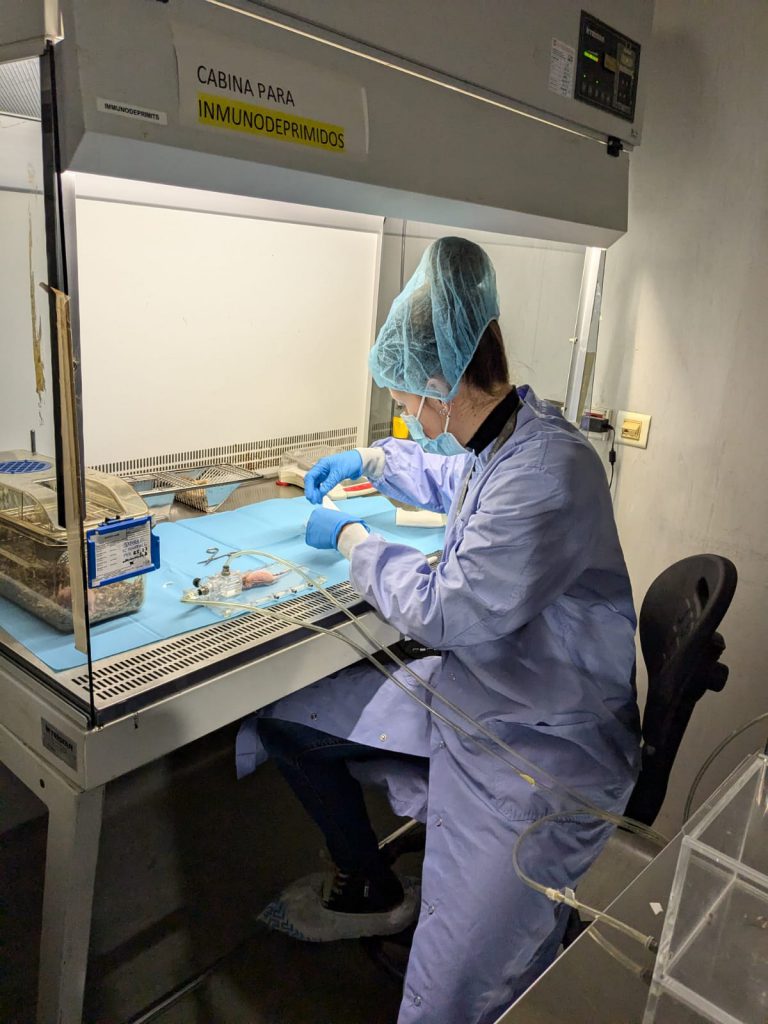
JUMISC Animal Housing Unit provides services to companies and research groups in multiple preclinical trials. This unit develops designs and performs preclinical validation on demand. It counts with professionals qualified to design, monitor and validate any type of biomedical study. An important feature of this unit is the possibility to house, maintain and supervise large animals for preclinical validation. This service also counts with a dilated experience in the handling and care of minipigs and what its use in preclinical studies implies.
It has a floor net area of 2.865 m2. It is equipped with spacious rooms to house experimentation animals depending on its specie characteristics (age, sex, weight, breed) and the research projects they are assigned to. Among the equipment and facilities are: rooms for the maintenance of large animals, rooms for the maintenance of small ruminants, rooms for the maintenance of birds, barrier-zone for the maintenance of rodents in SPF conditions, an operating room integrated within the barrier area, multipurpose rooms for the maintenance of rodents and lagomorphs, a room for the reception and handling of the animals, quarantine rooms for pre-experimental processes, a room for small procedures and cures and rooms for the preparation of large animals prior to the surgical area.

Among the services that the Unit develops, we highlight the following:

  • U22-S01 Housing and care of animal models.
  • U22-S02 In vivo experimental design/consultancy for preclinical procedures.
  • U22-S03 In vivo Toxicological studies in rodent and non-rodent species.
  • U22-S04 In vivo Biocompatibility studies of medical devices in rodent and non-rodent species.
  • U22-S05 PK/PD studies in rodent and non-rodent species.
  • U22-S06 In vivo Safety and efficacy studies of nanomaterials, biomaterials and new drugs in rodent and non-rodent species.
  • U22-S07 Development of animal models of disease in large animals.
  • U22-S08 Dosing of test substances and non-clinical specimen drawing in rodent and non-rodent species.

Customer benefits

All rooms and facilities included in the unit are certified with ISO-9001 and Good Laboratory Practices (GLP), strict quality standards that allow the production of highly accurate results.

In this way, preclinical, security and efficacy studies in animal models can be performed in compliance with the strict guidelines of the regulation agencies, ensuring reliability and traceability of all tests and results performed in its different services.

In addition, this team has been certified by the Spanish Agency of Drugs and Health Products (AEMPS) for studies of “Dosification of problem substances and non-clinical samples”, “Biocompatibility studies of health products”, “In vivo toxicity”, “Tolerance” and “Pharmacodynamics”. All these certifications allow to carry out studies to verify the efficacy, security and biocompatibility of nanotechnological development.

Target customer

The offered services can be of interest to a large number of companies, including the following:

Pharmaceutical and biotechnological companies: Companies that carry out preclinical and clinical studies for the development of drugs, therapies and biotechnological products and require animals to test the efficacy and security of their products.

Academic Researchers: Professors, researchers and students from academic institutions who need access to animals to conduct scientific research in areas like biology, medicine, behaviour, etc.

Medical Institutions: Hospitals, clinics and medical research centres that conduct studies to understand human diseases and develop medical treatments, including preclinical trials on animals.

Medical devices companies: Medical devices manufacturers who need to test the security and efficacy of their products on animals before commercialization.

Regulatory agencies: Government agencies in charge of regulating and monitoring security and efficacy of medical and pharmaceutical products, which may require data from animal studies to support products approval.

Food and agriculture companies: Companies that develop food and agricultural techniques may need to conduct animal studies to evaluate food security, the effects of additives, efficacy in agricultural products, etc.

Additional information

Selected publications:

  • Peiro JL, et al. Fetal Endoscopic Third Ventriculostomy Is Technically Feasible in Prenatally Induced Hydrocephalus Ovine Model. Neurosurgery 2023; Jun 1;92(6): 1303-1311.
  • Blanco-Blázquez V, et al. Swine Models of Aneurysmal Diseases for Training and Research. J Vis Exp 2022; Mar 23: 181.
  • Lucas-Cava V, et al. Prostatic artery occlusion versus prostatic artery embolisation for the management of benign prostatic hyperplasia: early results in a canine model. British Journal of Radiology 2022; 95: 1136.
  • Soria F, et al. Assessment of a Coated Mitomycin-Releasing Biodegradable Ureteral Stent as an Adjuvant Therapy in Upper Urothelial Carcinoma: A Comparative In Vitro Study. Polymers 2022; 14: 3059.
  • Campos JL, et al. Popliteal Vascular Lymph Node Resection in the Rabbit Hindlimb for Secondary Lymphedema Induction. J Vis Exp 2022; Nov 30: 189.
  • de la Cruz JE, et al. Biodegradable ureteral stents: in vitro assessment of the degradation rates of braided synthetic polymers and copolymers. Am J Clin Exp Urol 2022; Feb 15;10(1) :1-12.
  • Crisóstomo V, et al. The epicardial delivery of cardiosphere derived cells or their extracellular vesicles is safe but of limited value in experimental infarction. Sci Rep 2021; Nov 12;11(1): 22155.
  • Ballestín A, et al. A Pre-clinical Rat Model for the Study of Ischemia-reperfusion Injury in Reconstructive Microsurgery. J Vis Exp 2019; Nov 8: 153.
  • López E, et al. Identification of very early inflammatory markers in a porcine myocardial infarction model. BMC Vet Res 2019; Mar 12;15(1): 91.
  • Enciso S, et al. Validation of a model of intensive training in digestive laparoscopic surgery. Cir Esp 2016; Feb;94(2):70-6.
  • Crisostomo V, et al. Allogeneic cardiac stem cell administration for acute myocardial infarction. Expert Rev Cardiovasc Ther 2015; 13(3): 285-99.
Read More

U18-S01. In vivo nanotoxicology

In vivo nanotoxicology

The aim of this service is to evaluate the in vivo toxicity of nanoparticles and drug-loaded nanoparticles. Thus, we offer different mouse models in which we can determine the toxicity of the compounds. We offer to evaluate the nanotoxicology in healthy mice of any strain or in tumor mouse models. In the service we have subcutaneous and orthotopic/disseminated models of lymphoma, leukemia and colorectal, endometrium, head and neck carcinomas. Moreover, we have also available PDX models of colorectal and endometrium cancer. We can also offer the possibility to set up new animal models of other tumor types.

Customer benefits

The customer will benefit from the experience of the researchers involved in the service that have evaluated the nanotoxicology of a high number of diverse nanoparticles. The service has also high expertise in developing new cancer animal models. Thus, we offer also the possibility of setting up new models of any cancer type that fit with the needs of the customer. Moreover, the conditions of the assays are flexible and will be adapted to the need of each specific compound.

Target customer

The offered service can be of interest to research groups of academia or companies willing to test the nanotoxicology of nanoparticles or drug-loaded nanoparticles in vivo.

References

  • Martínez-Torró C, Alba-Castellón L, Carrasco-Díaz LM, Serna N, Imedio L, Gallardo A, Casanova I, Unzueta U, Vázquez E, Mangues R, Villaverde A. Lymphocyte infiltration and antitumoral effect promoted by cytotoxic inflammatory proteins formulated as self-assembling, protein-only nanoparticles. Biomed Pharmacother. 2023 Aug;164:114976. doi: 10.1016/j.biopha.2023.114976.
  • Rioja-Blanco E, Arroyo-Solera I, Álamo P, Casanova I, Gallardo A, Unzueta U, Serna N, Sánchez-García L, Quer M, Villaverde A, Vázquez E, Mangues R, Alba-Castellón L, León X. Self-assembling protein nanocarrier for selective delivery of cytotoxic polypeptides to CXCR4+ head and neck squamous cell carcinoma tumors. Acta Pharm Sin B. 2022 May;12(5):2578-2591. doi: 10.1016/j.apsb.2021.09.030.
  • Falgàs A, Garcia-León A, Núñez Y, Serna N, Sánchez-Garcia L, Unzueta U, Voltà-Durán E, Aragó M, Álamo P, Alba-Castellón L, Sierra J, Gallardo A, Villaverde A, Vázquez E, Mangues R, Casanova I. A diphtheria toxin-based nanoparticle achieves specific cytotoxic effect on CXCR4+ lymphoma cells without toxicity in immunocompromised and immunocompetent mice. Biomed Pharmacother. 2022 Jun;150:112940. doi: 10.1016/j.biopha.2022.112940.
  • -Medina-Gutiérrez E, García-León A, Gallardo A, Álamo P, Alba-Castellón L, Unzueta U, Villaverde A, Vázquez E, Casanova I, Mangues R. Potent Anticancer Activity of CXCR4-Targeted Nanostructured Toxins in Aggressive Endometrial Cancer Models. Cancers (Basel). 2022 Dec 23;15(1):85. doi: 10.3390/cancers15010085.
  • Sala R, Rioja-Blanco E, Serna N, Sánchez-García L, Álamo P, Alba-Castellón L, Casanova I, López-Pousa A, Unzueta U, Céspedes MV, Vázquez E, Villaverde A, Mangues R. GSDMD-dependent pyroptotic induction by a multivalent CXCR4-targeted nanotoxin blocks colorectal cancer metastases. Drug Deliv. 2022 Dec;29(1):1384-1397. doi: 10.1080/10717544.2022.2069302.
  • Serna N, Falgàs A, García-León A, Unzueta U, Núñez Y, Sánchez-Chardi A, Martínez-Torró C, Mangues R, Vazquez E, Casanova I, Villaverde A. Time-Prolonged Release of Tumor-Targeted Protein-MMAE Nanoconjugates from Implantable Hybrid Materials. Pharmaceutics. 2022 Jan 14;14(1):192. doi: 10.3390/pharmaceutics14010192.
  • Pallarès V, Unzueta U, Falgàs A, Aviñó A, Núñez Y, García-León A, Sánchez-García L, Serna N, Gallardo A, Alba-Castellón L, Álamo P, Sierra J, Cedó L, Eritja R, Villaverde A, Vázquez E, Casanova I, Mangues R. A multivalent Ara-C-prodrug nanoconjugate achieves selective ablation of leukemic cells in an acute myeloid leukemia mouse model. Biomaterials. 2022 Jan;280:121258. doi: 10.1016/j.biomaterials.2021.121258.
Read More

U17-S01. Confocal Microscopy Service

Confocal Microscopy Service

The Confocal Microscopy Service offers the possibility of making optical section of samples and enabling their three-dimensional reconstruction, which allows their visualization in high resolution with exceptional clarity and detail.
The unit is equipped with a Leica TCS-SP5 confocal microscope. The confocal module is equipped with three spectral detection channels, AOBS (Acousto-optical beam splitter) and a resonant scanner system that allows analysis at high speed and resolution and makes possible the analysis of dynamic in vivo physiological processes in short periods of time, significantly improving the survival of living biological samples by shortening the exposure times to lasers. Includes 1 argon laser, 1 He/Ne laser, 1 DPSS laser diode and 1 violet excitation laser. The microscope is coupled to a cell incubation kit that allows multi-position time lapse experiments. The equipment includes a workstation and four software for acquisition and analysis, which allow 3D visualizations, co-location studies, FRAP (Fluorescent Recovery after Photo-bleaching), FLIP (Fluorescent Loss in Photobleaching) and FRET (Fluorescence Resonant Energy Transfer). The equipment allows 3D characterization in detail of living cells and tissues through the use of different fluorochromes, expression and localization of molecules in 2/3D, colocalization and interaction of proteins or other types of molecules; endocytosis and intracellular transport, in situ hybridization with fluorescent probes, interaction studies between cells and materials, etc.
The unit provides researchers with a wide array of routine and specialized services as well as the latest advances in microscopy, including technical and scientific support to scientists for the study of cell/tissue biology, physiology and pathogenesis of diseases. Overall, the Confocal Microscopy Service serves as a valuable tool to advance scientific research and understand complex biological systems at the cellular and molecular level.

Customer benefits

Confocal microscopy is useful in biomedical science, cell and molecular biology and physiology for visualizing subcellular organelles, cellular interactions, disease mechanisms, co-localization studies, analysis of gene expression or in vivo and real-time experiments using markers or fluorescent fusion proteins.
Also, in tissue engineering and materials science, confocal microscopy enables surface morphology characterization, and material properties.

Confocal Microscopy Service offers specific advantages to customers by providing high-resolution imaging and adding value through access to advanced instrumentation and expertise. It becomes essential for organizations involved in Research and Development across various sectors, enabling them to advance scientific understanding, accelerate innovation, and drive discoveries that have social impact.

Target customer

The Confocal Microscopy Service becomes essential for organizations involved in Research and Development in various scenarios:

  • Biomedical Research Institutes: This service can be useful for research institutions focused on the study of cellular and molecular mechanisms underlying different diseases, allowing to visualize pathological changes, identify biomarkers and investigate possible therapeutic targets. Additionally, it allows researchers to study disease progression, evaluate drug efficacy, and develop new treatment strategies.
  • Pharmaceutical Companies: Confocal microscopy can be used by drug development companies to evaluate the effects of these drugs on cellular function.
  • Academic Laboratories: Confocal microscopy can be useful for researchers in diverse fields of biology, including neuroscience, developmental biology, and cancer biology, to explore fundamental biological processes and generate novel scientific insights.

References

  1. Benito-Martínez S, Pérez-Köhler B, Rodríguez M, García-Moreno F, Gómez-Gil V, Pascual G, Bellón JM. Antibacterial Biopolymer Gel Coating on Meshes Used for Abdominal Hernia Repair Promotes Effective Wound Repair in the Presence of Infection. Polymers (Basel). 2021;13(14):2371. doi: 10.3390/polym13142371.
  2. Sánchez-Esteban S, Castro-Pinto M, Cook-Calvete A, Reventún P, Delgado-Marín M, Benito-Manzanaro L, Hernandez I, López-Menendez J, Zamorano JL, Zaragoza C, Saura M. Integrin-Linked Kinase Expression in Human Valve Endothelial Cells Plays a Protective Role in Calcific Aortic Valve Disease. Antioxidants (Basel). 2022. 11(9):1736. doi: 10.3390/antiox11091736. 
  3. Campillo S, Bohorquez L, Gutiérrez-Calabrés E, García-Ayuso D, Miguel V, Griera M, Calle Y, de Frutos S, Rodríguez-Puyol M, Rodríguez-Puyol D, Calleros L. Indoxyl sulfate- and P-cresol-induced monocyte adhesion and migration is mediated by integrin-linked kinase-dependent podosome formation. Exp Mol Med. 2022;54(3):226-238. doi: 10.1038/s12276-022-00738-8. 
Read More

U16-S01. X-ray photoelectron spectroscopy service

X-ray photoelectron spectroscopy service

This service provides quantitative surface chemical elemental composition of a wide range of materials by X-ray photoelectron spectroscopy (XPS). In addition to obtaining the elemental composition of the surface of the materials studied, it is possible to determine their chemical state and the overall electronic structure and density of electronic states of the material. Using this technique, it is possible to check which elements are present on the surface of the material, as well as to which other elements they are bonded. In addition, this service offers the possibility of depth profiling when combined with ion beam etching.

Customer benefits

The instruments are located in the Research Support Services, specifically in the Analysis and Characterisation of Solids and Surfaces Service. Within the service there is a Doctor responsible for the instruments, in charge of their management and handling, as well as optimising their use as much as possible.

Target customer

Being located within the Research Support Services, it allows full access to the instruments (handled by a specialist in the technique) to the entire scientific community, both at the University of Extremadura and other public and private organisations.

Additional information

C:\Users\usuario\Desktop\Proyectos\CIBER\Nanbiosis\Fotos XPS\1677655978522.jpg

Read More

U16-S04. Detection of secondary ions by time-of-flight mass spectrometry service (TOF-SIMS)

Detection of secondary ions by time-of-flight mass spectrometry service (TOF-SIMS)

This service employs one of the most powerful techniques at the level of detection and resolution both laterally and in depth for chemical composition measurements on surfaces and depth on thin films, such as TOF-SIMS or detection of secondary ions by time-of-flight mass spectrometry. In this way, the chemical composition of different materials can be tracked to form 2D and 3D conformational depth mappings or profiles of the system under study. In addition, the service allows surface analysis of organic and inorganic materials by obtaining mass spectra, mapping of chemical elements present on the surface of the sample by imaging and depth profile analysis.

Customer benefits

The instruments are located in the Research Support Services, specifically in the Analysis and Characterisation of Solids and Surfaces Service. Within the service there is a Doctor responsible for the instruments, in charge of their management and handling, as well as optimising their use as much as possible.

Target customer

Being located within the Research Support Services, it allows full access to the instruments (handled by a specialist in the technique) to the entire scientific community, both at the University of Extremadura and other public and private organisations.

Additional information

C:\Users\usuario\Desktop\Proyectos\CIBER\Nanbiosis\Fotos TOF\TOF-SIMS 1.jpg

Read More

U15-S01. Magnetometry (Remote) OUTSTANDING

Magnetometry on nanoparticles in solid form, ultra thin films, powders, liquids and even slurries:

  • Magnetization curves.
  • Coercitivity (normal and remanent).
  • Magnetization vs. time curves.
  • First Order Reversal Curves (FORC) diagrams.
  • Diamagnetic and paramagnetic susceptibility.
  • S* (measurement of the gradient in the second quadrant).
  • Remanent and saturation magnetization.
  • Initial permeability.

Measurements of magnetic properties of materials:

  • Diamagnetic, paramagnetic and ferromagnetic materials.
  • Magnetic recording media.
  • Magnetoresistive Random-Access Memory (MRAM).
  • Amorphous metals.
  • Giant Magnetoresistance Effect (GMR).

Geophysical Research:

  • Measurements of magnetization of rock, sediment and organic samples.

Biomedical Research:

  • Detection of small nanoparticles inside biological tissue in order to study the biodistribution and toxicity.
  • Study of the iron levels which are associated with some types of neurodegenerative disease.
Read More

U15-S02. Relaxometry (Remote) OUTSTANDING

Measurement of the relaxation times in aqueous solutions and biological samples containing superparamagnetic nanoparticles as contrast agents for MR images. For T1 in the continuous range 10 kHz to 80MHz; for T2 in the range 10 MHz to 80MHz.

Characterization of contrast agents for MR imaging, ascertaining their relaxivity and the dominant effect.

NMR relaxometry technique is an important analytical tool for NMR research and material characterization in both industrial and academic environments and has been successfully applied in a wide range of fields:

– Pharmaceutical Applications:

  • R&D of MRI contrast agents
  • Proteinstudies
  • R&D for formulations (e.g. properties of solutions; liposome carriers)
  • Quality control of products in manufacturing

– Polymers Applications:

  • R&D for new polymer materials
  • Control of levels of polymer additive
  • Quality control of products in manufacturing

– Oil, gas and petroleum applications:

  • Oil and gas surveying – rock pore size evaluation
Read More