+34 620 10 75 37info@nanbiosis.com

Preclinical validation bioimaging - Equipment

Preclinical validation bioimaging

U23-09 Time Lapse Incubator MIRI TL

The MIRI® Time-Lapse Incubator is a multiroom incubator with a built-in camera and microscope. The MIRI® Time-Lapse Incubator provides high quality time-lapse images of embryos developing in “real-time” without having to remove the embryos from the safety of the incubation chamber for manual microscopy. Time-lapse embryo monitoring provides detailed morphokinetic data throughout embryo development, which is not available on routine spot microscopic evaluation. This allows all important events to be observed, helping to identify healthy embryos with the highest probability of implantation, with the aim of achieving higher pregnancy rates. Additionally, each individual incubator has its own CO2 and oxygen temperature control. which makes them not interfere with each other.


Read More

U23-07 PiezoXpert (Piezoelectric micromanipulation and microinjection).

This equipment facilitates the piezo-assisted microinjection of sperm into the cytoplasm of eggs from different animal species (mouse, horse, sheep, porcine, etc.) that could be damaged by conventional ICSI given their elasticity, for this purpose and through the piezoelectric technique that It transforms electrical pulses into mechanical ones, the cell membrane is perforated with effectiveness and reproducibility, allowing the sperm to enter the cytoplasm of the oocyte without damaging it, improving the results of ICSI in animal models. Furthermore, this technique can be used in many processes such as the production of transgenic animals, IVF, developmental biology, and other applications in cell biology.

Read More

U26-E07. MRS*DRYMAG3017-Flexiscan

Equipment Features:

Description: 3T preclinical imaging equipment with PET ClipOn attached.

Technical specifications: Imaging equipment with superconducting magnet, capable of varying the angle from 0 to 90º, without cryogenics of 3 Teslas; mouth diameter of 170mm; 600 mT/m gradients with 5% linearity; 30 mm DSV for Homogeneity and 0.05 ppm/hour for stability; 350 Kg of total weight.

Applications:

Acquisition and viewing of in vivo images of mice and rats as well as other beings or materials of similar size.

  • Qualitative and quantitative studies (relaxation times T1 and T2 and even making maps) at the brain level and other organs.
  • Cardiological sequences: diffusion, determination of blood volume…
    Anatomical and functional studies of different organs (for example: renal, abdominal, liver…).
  • Bone-level imaging, distinguish between bone material from soft tissue.
    Fat suppression techniques.
  • Oncological studies to detect and locate tumors and volume calculation.
  • Carry out studies on fruits and vegetables (grapes, kiwi, tomato, seeds…).
  • Perform studies with/without contrast (Gadolinium) and see the differences between the 2 images.
  • Thanks to the possibility of rotating the equipment in a vertical position, images of plants can be obtained (because by their nature, they have to be in a vertical orientation, such as a stem).
  • Ex-vivo studies can also be carried out vertically and in this way we maintain the integrity of the organ (eg heart).
Read More

U22-E09. Metabolic cages to perform toxicity studies in rodents

Metabolic cages to perform toxicity studies in rodents

Read More

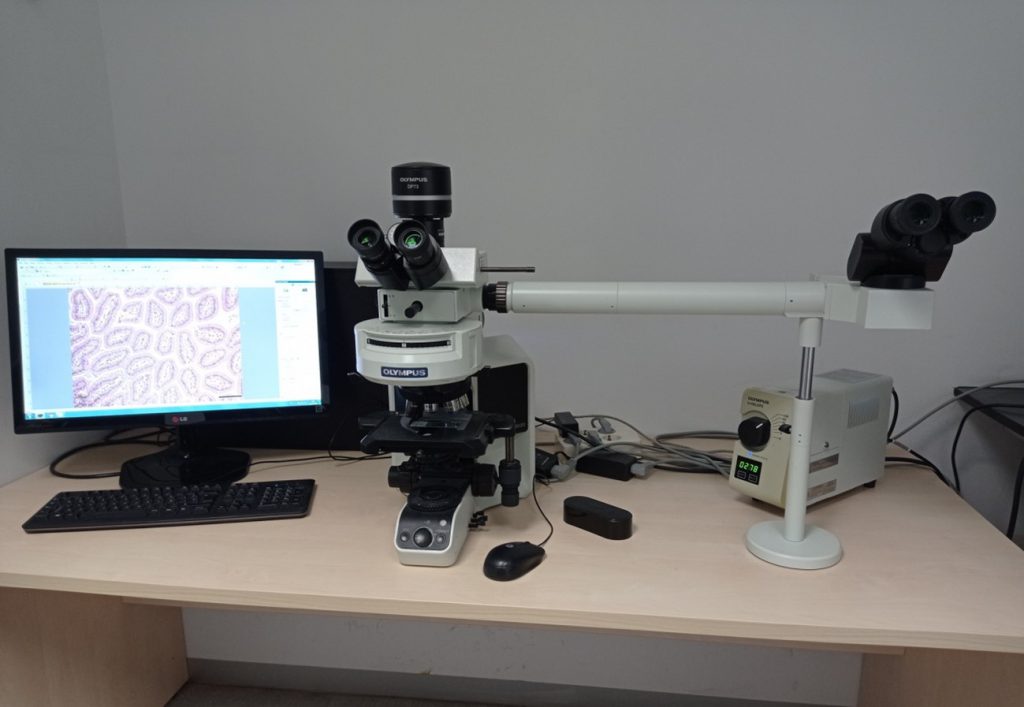
U18-E13. Immunofluorescenece supplement for Olympus Microscope

Immunofluorescenece supplement for Olympus Microscope for immunofuorescent evaluaction of cells and tissues


Read More

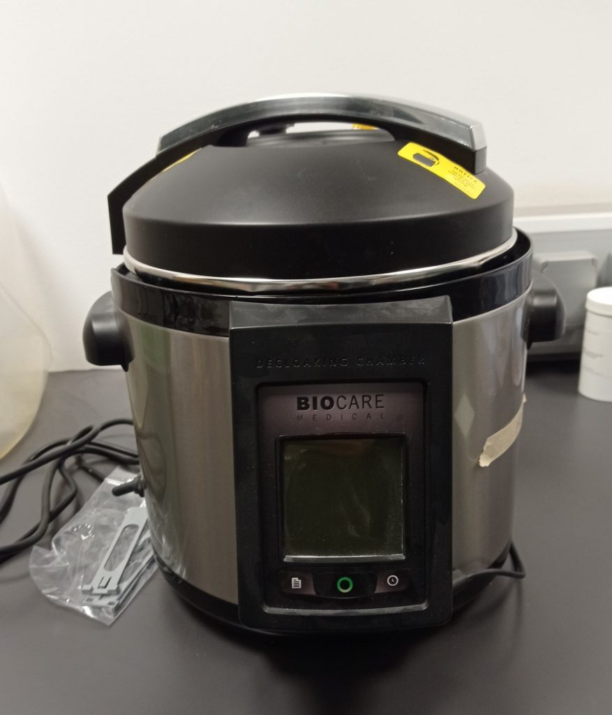
U18-E12. Decloaking Chamber NxGen (Anacron diagnostics)

Decloaking Chamber NxGen (Anacron diagnostics); Charmber for antigen retrieval for immunohistochemical assays

Read More

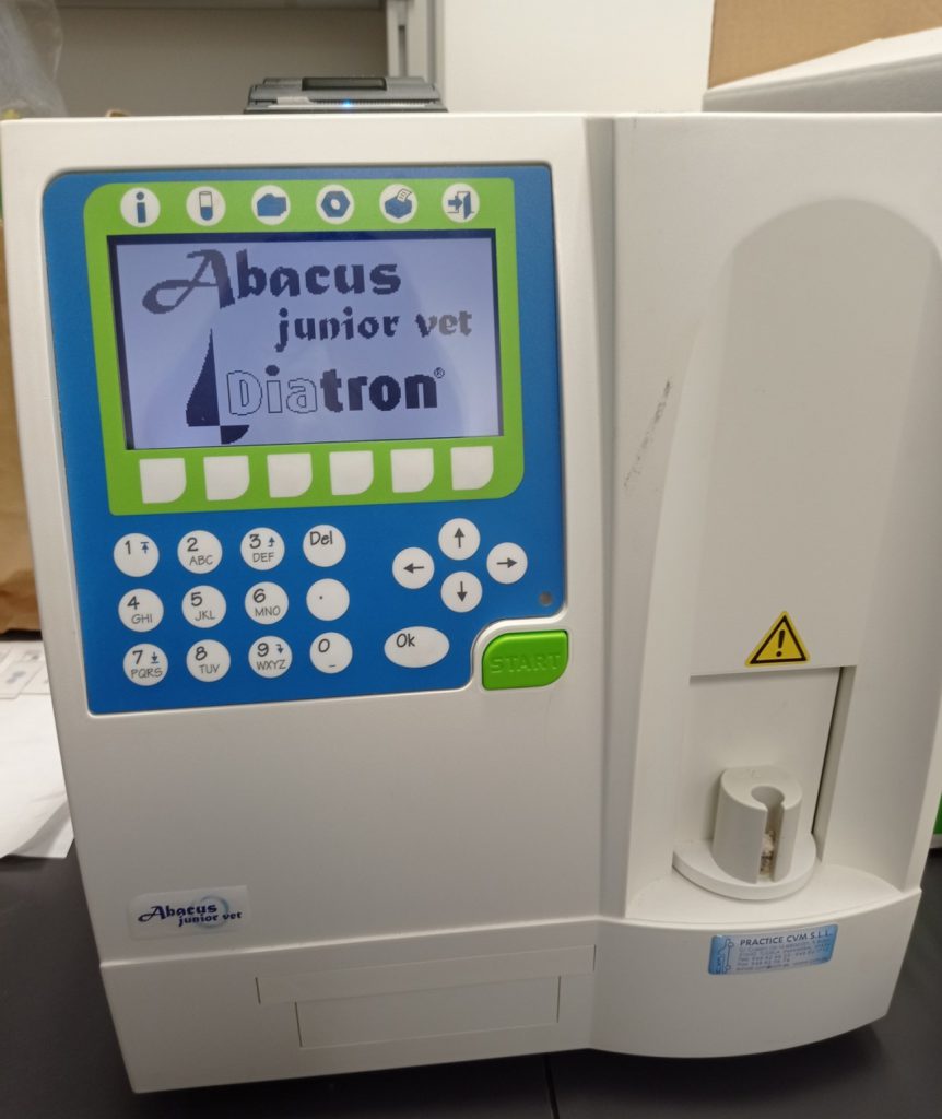
U18-E11. Abacus hematocytometer

Abacus hematocytometer to determine the leucocytary formula in animal blood.


Read More

U20-E04. General laboratory equipment

General laboratory equipment

Read More

U26-E06. Gilson 215 liquid handler for automated sample preparation of liquid samples

Read More

U26-E04. Bruker DPX300 spectrometer(7 T) with standard entrance, and two RF channels.

Accessories:

  • 5mm QNP (1H, 13C, 31P and 19F), with pneumatic accessory for automatic nucleus change and field gradients along the z axis.
  • BACS autosampler for 60 samples.
Read More