+34 620 10 75 37info@nanbiosis.com

News U25

MRSI-based generation of nosological images for noninvasive therapy response assessment in preclinical GBM

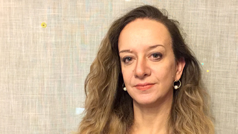
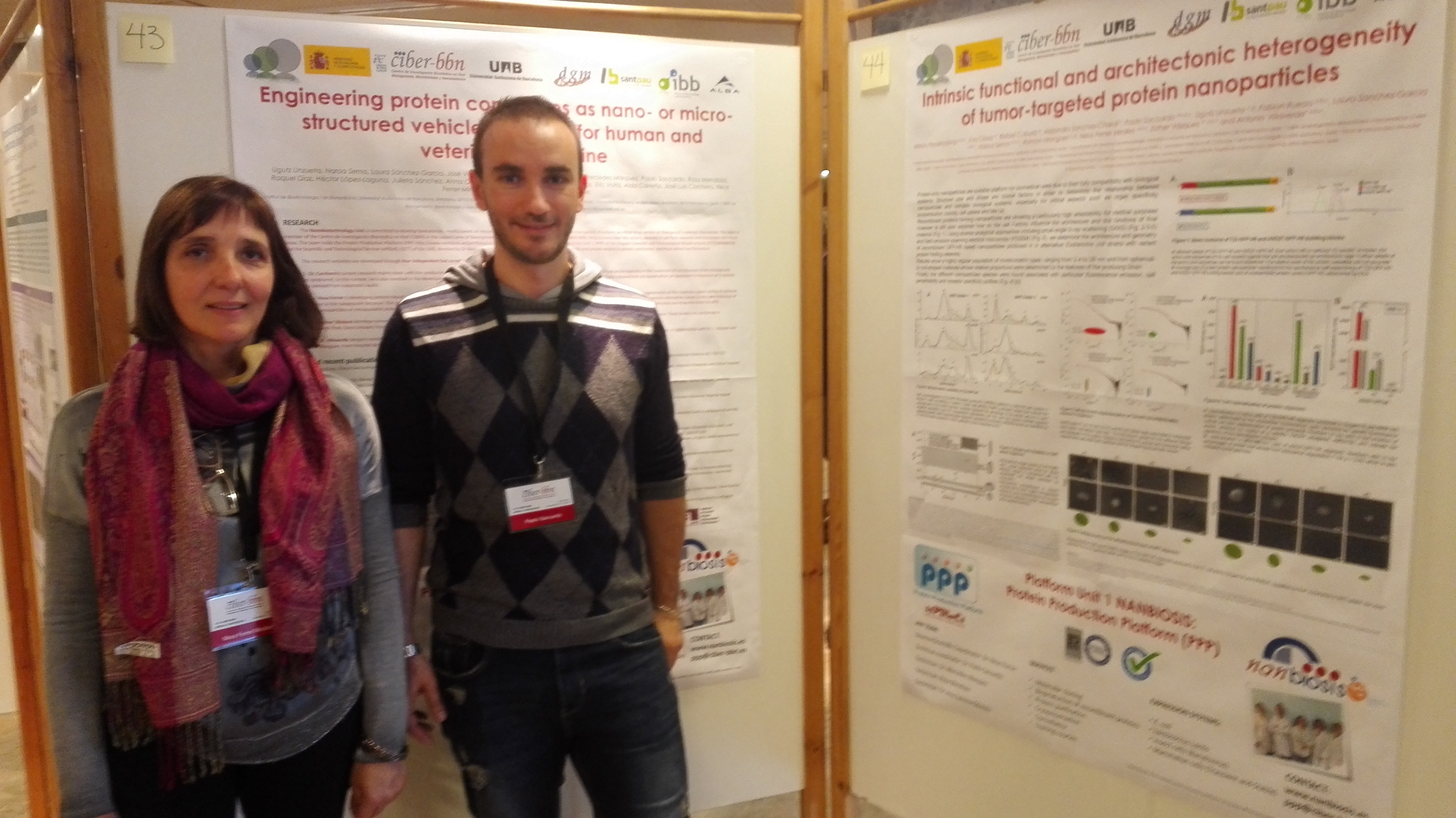
Nuria Arias Ramos from GABRMN-UAB, research group coordinator of Unit 25 of NANBIOSIS, presented on January 30th, an oral communication at the youngSMIN | young Spanish Molecular Imaging Network Meeting.

The title of the communication is “Multi slice MRSI-based generation of nosological images for noninvasive therapy response assessment in preclinical GBM”, written by Nuria Arias-Ramos, SilviaLope-Piedrafita, Victor Mocioiu, Margarida Julià-Sapé, Carles Arús and Ana Paula Candiota.

The work is basically centered in extending the preliminary GABRMN study based on MRSI  acquired in a preclinical glioblastoma (GB), both under treatment and control, with pattern recognition and source extraction for noninvasive therapy response assessment. The preliminary study was  performed in a single tumoral slice, but the GB is a well-known heterogeneous tumor and also presents heterogenous response to treatment, so the multislice acquisition was an important step in order to characterize therapy response assessment as a whole, not centered in a single slice. Tumors were stratified in no response, partial response and high response after calculating the percentage of the ‘responding’ part of the tumor, and histopathological studies for correlation are in progress.

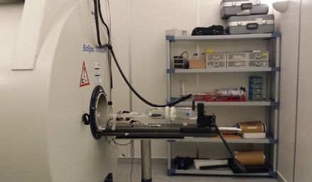
The MRSI acquisition and processing steps were performed in NANBIOSIS facility U25.

MRSI-based generation of nosological images for noninvasive therapy response assessment in preclinical GBM
Read More

NANBIOSIS lecturers in B-Debate Imaging for Life. From Molecules to Diagnostics and Therapy

Last November took place in CosmoCaixa, Barcelona, the B-Debate Days Imaging for Life. Molecules from Diagnostics and Therapy.

The event, organized by B·Debate (an initiative of Biocat and “la Caixa” Foundation), IBB and LJS, was focussed on Biomolecular Imaging, with emphasis on molecular, cellular, tissular and small animal research, as well as biomedical applications, including noninvasive diagnostics, image guided surgery, multimodal imaging and theranostics.

Scientists of NANBIOIS Unit 20 and 25 were invited to participate as guest lecturers:

-Simó Schwartz, Scientific Director of Unit 20: New bioluminescent models to target cancer stem cells with nanomedicine.

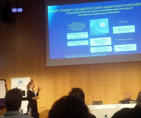
-Ana Paula Candiota, Scientific Coordinator of Unit 25: Unravelling therapy response in preclinical glioblastoma using MRSI-based molecular imaging and source analysis.

As Professor Candiota explained “Characterization of Glioblastoma (GB) response to treatment is a key factor for improving patient survival and prognosis. Magnetic Resonance Imaging and Spectroscopic Imaging (MRI/MRSI) provide morphologic and metabolic profiles of GB but usually fail to produce unequivocal surrogate biomarkers of response. Ideally, we would like to provide clinicians with early therapy response follow-up and an improved time frame for changing or adapting therapy schemes”. Her talk focussed in the capability of advanced pattern recognition techniques, such as semi-supervised signal source extraction, to produce nosological images with robust recognition of response to temozolomide (TMZ) in preclinical GB (GL261 tumour-bearing in immunocompetent C57BL/6 mice) through the information contained in MRSI grids. These techniques have a clear translational potential and could improve future patient management and care. The acquisition of mice MRSI data used for pattern recognition strategies development and evaluation was performed in the Biospec 7T scanner from U25.

NANBIOSIS lecturers in B-Debate Imaging for Life. From Molecules to Diagnostics and Therapy
Read More