+34 620 10 75 37info@nanbiosis.com

News

nanbiosis news

New patented peptide to allows the faster internalization of drugs within cells and the design of more effective therapeutic nanoconjugates

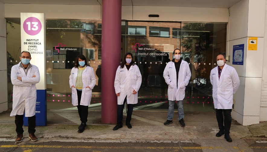
Researchers of NANBIOSIS Unit 20 In vivo Experimental Platform of CIBER-BBN and Vall d’Hebron Research Institute (VHIR) have patented a peptide that, in comparison to the current standard treatment, is much faster, internalizes much more, and does not cause any toxicity.

The membrane of a cell is an effective barrier that hinders the targeted delivery of molecules, such as therapeutic compounds. During the last years, several strategies have been developed to get the molecules into the cell interior but, in general, the methods developed still show a low efficacy and / or toxicity. “The use of therapeutic nanoconjugates such as nanomedicines facilitates the transport and delivery of drugs in target cells, but often with less efficiency than we would like,” says Dr Simó Schwart Jr, head of the Scientific Director of NANBIOSIS Unit 20 and the CIBBIM-Nanomedicine group: Direction i Alliberament Farmacològic del Vall d’Hebron Research Institute (VHIR)/CIBER-BBN.

Given the need to get more drugs or proteins into cells, one of the alternatives to be able to increase the amount that enters their interior more quickly is what is known as Cell penetrating peptides or cellular internalizing peptides, small sequences of amino acids that have the ability to interact with the plasma membranes of cells and, as a result of this interaction, make it easier to internalize the cargo they carry. An example of application would be when an internalizing peptide binds to a therapeutic nanoconjugate, achieving a greater capacity for the nanoconjugate to enter the cell interior and, therefore, to release the drugs it carries into the cells.

Until now, one of the most important internalizing peptides used has been known as TAT. Now, a team of researchers led by Dr. Schwartz Jr, has discovered a sequence common to a family of peptides that significantly outperforms the TAT results and facilitates the cellular internalization of nanoconjugates in a very significant way. These peptides are derived from a membrane protein called CD300 which has a very high capacity to interact with sphingomyelin, a lipid found in all plasma membranes and also in intracellular organelles. “The peptides in our patent”, explains Dr. Simó Schwartz Jr, “are derived from an extracellular part of CD300, which has a high capacity to bind sphingomyelin. Compared to the current standard treatment, TAT, CD300f7 is much faster, internalizes much more, and does not cause any toxicity. The use of these peptides in nanomedicine therefore facilitates and increases the internalization process of all the cargo they carry. This means that we are able to introduce drugs into cells in less time and in greater quantities ”. The results of this discovery not only allow for faster internalization within the cell, but also open the door to designing much more effective therapeutic nanoconjugates.

Souce of information: VHIR news

Read More

Graphene active sensor arrays for long-term and wireless mapping of wide frequency band epicortical brain activity

Researchers of Nanbiosis U8 Micro– Nano Technology Unit, from CIBER-BBN and IMB-CNM-CSCIC have published an article in Nature Communications on Graphene arrays for long-term and wireless mapping of epicortical brain activity. A collaborative work in the framework of the Brain Com and Graphene EU projects. The article mentions the participation of NANBIOSIS-ICTS.

Graphene active sensors have demonstrated promising capabilities for the detection of electrophysiological signals in the brain. Their functional properties, together with their flexibility as well as their expected stability and biocompatibility have raised them as a promising building block for large-scale sensing neural interfaces. However, in order to provide reliable tools for neuroscience and biomedical engineering applications, the maturity of this technology must be thoroughly studied. Here, we evaluate the performance of 64-channel graphene sensor arrays in terms of homogeneity, sensitivity and stability using a wireless, quasi-commercial headstage and demonstrate the biocompatibility of epicortical graphene chronic implants. Furthermore, to illustrate the potential of the technology to detect cortical signals from infra-slow to high-gamma frequency bands, we perform proof-of-concept long-term wireless recording in a freely behaving rodent. Our work demonstrates the maturity of the graphene-based technology, which represents a promising candidate for chronic, wide frequency band neural sensing interfaces.

Article:

Garcia-Cortadella, R., Schwesig, G., Jeschke, C. et al. Graphene active sensor arrays for long-term and wireless mapping of wide frequency band epicortical brain activity. Nat Commun 12, 211 (2021). https://www.nature.com/articles/s41467-020-20546-w

Read More

A rare genetic bone pathology is identified from massive sequencing methods

The Andalusian Center for Nanomedicine and Biotechnology (BIONAND) and the Center for Biomedical Research Network in Bioengineering, Biomaterials and Nanomedicine (CIBER-BBN), both partners of NANBIOSIS, in collaboration with the International Registry of Skeletal Dysplasias of the University of California (Los Angeles) and Masaryk University, of the Czech Republic have described a new genetic disease of the skeleton using a precision medicine strategy.

This disease consists of extreme bone fragility with lack of mineralization and skeletal deformation associated with joint dislocation and heart disease, as well as a lung deficiency that causes perinatal lethality -at the time of birth-. Using massive sequencing methods – of all genes – researchers have identified the mutations that caused a type of rare bone pathology, specifically, those of the ‘LAMA5’ gene, responsible for encoding a cellular matrix protein that surrounds blood vessels in skeletal tissues.

Our scientific team has spent years investigating rare genetic syndromes that affect the skeleton in order to provide a medical solution to patients with difficult diagnosis and treatment,” explains the researcher from the Department of Cell Biology, Iván Durán, lead author of this study, whose results have been published in the scientific journal ‘EBIOMEDICiNE’.

According to the expert, precision medicine is the key to discovering what genetic and molecular factors cause this type of pathology and, therefore, understanding the mechanism that causes them and being able to develop personalized therapies.

Thus, researchers have also described the disease mechanism by generating cellular models by gene editing, mimicking the mutations in ‘LAMA5’, with the aim of confirming whether these are the origin and knowing the molecular process that triggers the problem. These cellular models have been generated by genetic editing with CRISPR, introducing mutations that cause a null or hypomorphic gene.

“Thanks to these models, we discovered a new signaling pathway that governs the formation of the skeleton – so that the bone grows and remains healthy – which means that our work has not only led to the discovery of a new disease, but to a mechanism unprecedented that can be exploited for common bone disorders ” –explains Durán, “the presence of ‘LAMA5’ between cells that direct skeletal formation indicates, therefore, that the appearance of signals from special blood vessels can be a very effective weapon for bone repair and regeneration. Blood vessels not only provide irrigation to the bone, but also carry signals and house niches of stem cells that can be mobilized to induce a regenerative process. ‘LAMA5’ seems to be a key component for harboring pericyte-type stem cells”.

Article of reference:

Barad M, Csukasi F, Kunova-Bosakova M, Martin J, Zhang W, Taylor SP, Dix P, Lachman R, Zieba J, Bamshad M, Nickerson D, Chong JX, Cohn DH, Krejci P, Krakow D, Duran I. Mutations in LAMA5 disrupts a skeletal noncanonical focal adhesion pathway and produces a distinct bent bone dysplasia. 2020 EBioMedicine. Nov 23;62:103075. doi: 10.1016/j.ebiom.2020.103075

Read More

Inkjet Priting Technology, Manufacture and validation of electrochemical sensors in medical applications

Miguel Zea, a member of the NANBIOSIS U8 Micro– Nano Technology Unit presents a video explaining his research is based on the manufacture and validation of electrochemical sensors in medical applications: –“Using InkJet printing I have made sensors in different plastics and paper. Also using a novel approach in each sensor. I have made two pH sensors using novel Platinum and polymer inks and also a cortisol sensor on paper”.

With this video, Miguel Zea, participates in the second edition of ‘I investigate, I am CSIC’. It is a competition hold by The Spanish National Research Council (CSIC) for its doctoral students to disseminate their doctoral thesis. Through short videos of maximum duration of 3 minutes, predoctoral scientists explain their research and results for the public in general

Here you can see the video and vote with a like!

Read More

Outstanding Young Researcher Award at ICESS 2021 to Konstantinos Mountris (NANBIOSIS U27).

Konstantinos Mountris researcher from the BSICoS group of CIBER-BBN and I3A at the University of Zaragoza has been granted the Outstanding Young Researcher Award at the International Conference on Computational & Experimental Engineering and Sciences (ICCES) in relation with the work Radial Point Interpolation Mixed Collocation (RPIMC) Method for The Solution of Reaction-Diffusion Equation in Cardiac Eletrophysiology (for the simulation of myocardial infarction).

This work was already recognized in the Congress of Computing in Cardiology (CinC) held recently where Konstantinos Mountris and Esther Pueyo have received the Maastricht Simulation Award (MSA)Konstantinos Mountris acknowledged the contribution of NANBIOSIS U27 High Performance Computing :“using the HPC services of NANBIOSIS U27 we were able to validate the RPIMC method as a promising alternative to Finite Element Method performing large-scale simulations of myocardial infarction in biventricular swine models

Related news: Understanding human heart behaviour with mathematics and engineering.

Read More

New Scanning Electron Microscope and on line Seminar to explain it for internal and external accesses on NANBIOSIS U12

A new Scanning Electron Microscope (Hitachi TM-4000 Plus II) has been installed at the Nanostructured liquid characterization unit of NANBIOSIS ICTS (Unit 12) of CIBER-BBN and IQAC-CSIC.

An online seminar will be given by Susana Vilchez on January 14th at 12h to explain the various features, functions and capabilities of this new instrument, Tabletop Scanning Electron Microscope Hitachi TM-4000 Plus II, that is open for both internal and external users.

Those interested in attending the seminar can contact unit 12 of NANBIOSIS:

Read More

NANBIOSIS Unit 2 obtains the Biosafety Level 2 Accreditation

The laboratory of NANBIOSIS Unit 2 from IQAC-CSIC and CIBER-BBN has obtained the biosafety level 2 accreditation, which allows the laboratory to work with biological agents classified in the risk group 2.

Biological containment level 2 (NBS2) laboratories are generally required to work with any derivative of human blood or other primates, body fluids (especially when they are visibly contaminated with blood), cell lines, or tissues in which has uncertainty about the presence of an infectious agent.

Also, the group participates in the COVID project “Point-of-care tests for the rapid detection of SARS-CoV-2 (POC4CoV)”, funded by the CSIC. This project involves the handling of swabs and serum samples from both positive and negative SARS-CoV-2 patients. Lluïsa Vilaplana, member of research group Nb4D of CIBER-BBN and IQAC-CSIC, led by Dra. M. Pilar Marco, wich Coordinates NANBIOSIS U2, has coordinated the process to obtain the accreditation.

The laboratory has an antechamber or clean locker room, a card-controlled entry system and a space for the storage of materials and reagents. It has also an independent air conditioning system, a specific air renewal system and a biosafety cabin type 2A, with HEPA filter. In addition, it is equipped with a suitable lighting system, an emergency lighting system and a large observation peephole on the door.

In addition to this specific equipment, the laboratory is equipped with an inverted microscope, centrifuge, thermostatic bath, stirrers, incubators, refrigerator and autoclave for sterilization and waste management.

The Nanobiotechnology for Diagnosis (Nb4D) research group , focus the research on the development of biomarkers for the diagnosis of infectious diseases. Nowadays, the group participates in five research projects related to this topic. These projects involve working with clinical samples for the detection of the pathogens Pseudomonas aeruginosa (Gram – type bacteria) and Staphylococcus aureus (Gram + type bacteria), both classified in risk group 2.

Source of information: IQAC-CSIC Communication

Read More

Chemical composition of explanted deteriorated nephrostomy polyurethane-catheters through X-ray photoelectron spectroscopy

Researchers of Nanbiosis U16 Surface Characterization and Calorimetry Unit of CIBER-BBN and University of Extremadura in Badajoz, are the authors of an article published by Materials Chemistry and Physics,

The researchers studied the surface chemical information of thirteen used catheters that had remained in patients for two months, using the XPS technique with the purpose of this communication is to report.
Compositional changes in relation to unused catheters provided information on the degree of the chemical degradation suffered. ATR-IR
added information on the chemical characterization of the samples
and Scanning Electron Microscopy (SEM) analysis will advise on topographical changes.

The XPS technique is optimum to analyzed the surface chemical composition of medical polymer device. Applied to the ureteral catheters, XPS shows on the surface of damaged catheters calcium and other ions from urine. XPS was performed by the ICTS “NANBIOSIS”, more specifically by the Surface Characterization and Calorimetry Unit of the CIBER in Bioengineering, Biomaterials & Nanomedicne (CIBERBBN)
and the SACSS-SAIUEx of the University of Extremadura (UEx)

Ureteral catheters are a fundamental part of the modern urologist’s armamentarium. X-ray photoelectron spectroscopy (XPS) was used for the first time as a powerful analytical tool for the study of the chemical
composition of nephrostomy catheters retrieved from patients who had undergone nephrostomy to detect their chemical deterioration inside the human body. Depth profile analysis provided not only the composition of the surface but also that of the catheter bulk. The results obtained by XPS showed the presence of calcium and other ions, such as phosphorus, sulphur and fluorine in the explanted deteriorated catheters. The detection of barium on the surface of all the retrieved catheters has special relevance. This chemical element is usually incorporated as a radiomarker in the catheter polymeric matrix and its diffusion from the bulk material to the surface must be responsible for its XPS detection. The accumulation of high levels of this element from toxic barium salts in cases of urine drainage failure could lead to its adsorption from the surrounding tissues into the patient’s body, thus compromising the safety concentrations of this soft alkaline earth metal.

Article of reference:

Chemical composition of explanted deteriorated nephrostomy polyurethane-catheters through X-ray photoelectron spectroscopy María Fernández-Grajera, Margarita Hierro-Oliva, Luis Fernández-deAlarcón, Amparo M.Gallardo-Moreno. Materials Chemistry and Physics Volume 239,  2020, 121979 https://doi.org/10.1016/j.matchemphys.2019.121979

Read More

Non-viral mediated gene therapy in human cystic fibrosis airway epithelial cells recovers chloride channel functionality

Researchers of CIBER-BBN Units of NANBIOSIS: U29 Oligonucleotide Synthesis Platform (OSP) at IQAC_CSIC, led by Prof. Ramón Eritja and U10 Drug Formulation, at UPV-EHU, led by Prof José Luis Pedraz, are coauthors of an article published by International Journal of Pharmaceutics.

Gene therapy strategies based on non-viral vectors are currently considered as a promising therapeutic option for the treatment of cystic fibrosis (CF), being liposomes the most commonly used gene carriers. Niosomes offer a powerful alternative to liposomes due to their higher stability and lower cytotoxicity, provided by their non-ionic surfactant and helper components. In this work, a three-formulation screening is performed, in terms of physicochemical and biological behavior, in CF patient derived airway epithelial cells. The most efficient niosome formulation reaches 28% of EGFP expressing live cells and follows caveolae-mediated endocytosis. Transfection with therapeutic cystic fibrosis transmembrane conductance regulator (CFTR) gene results in 5-fold increase of CFTR protein expression in transfected versus non-transfected cells, which leads to 1.5-fold increment of the chloride channel functionality. These findings highlight the relevance of niosome-based systems as an encouraging non-viral gene therapy platform with potential therapeutic benefits for CF.

The article acknowledges U10 Drug Formulation, for the intellectual and technical assistance

Article or reference:

Non-viral mediated gene therapy in human cystic fibrosis airway epithelial cells recovers chloride channel functionality-Sainz-Ramos, M., Villate-Beitia, I., Gallego, I., A.L. Qtaish, N., Lopez-Mendez, T.B., Eritja, R., Grijalvo, S., Puras, G., Pedraz, J.L. International Journal of Pharmaceutics, 588, art. no. 119757, 2020. https://doi.org/10.1016/j.ijpharm.2020.119757

Read More

New equipment for calorimetry and surface characterization for NANBIOSIS U16

NANBIOSIS unit 16 Surface Characterization and Calorimetry Unit form CIBER-BBN and University of Extremadura offers the performance of tasks of physical-chemical characterization of surfaces using techniques such as ellipsometry, calorimetry, X-ray photoelectronic spectroscopy (XPS) and detection of secondary ions by means of mass spectrometry by time of flight (Tof-SIMS). Recently, new equipment acquired through the execution an investment of 1.3 million euros, cofinanced with FEDER funds, the Ministry of Economy and Competitiveness and Junta de Extremadura, Regional Ministry of Economy, Science and Digital Agency, througth the project FICTS1420-14-09. These equipments are a microdroplet and picodroplet contact angle goniometry system and an optical profilometry system.

Equipment acquired

PEAK AND MICRO DROP GONIOMETRY SYSTEM

This system allows to measure contact angles automatically, controlling by software, the deposition of drops of different liquids, their analysis and the orientation of the substrate, as well as pending drops. In addition, the microdroplet system has a tilting base that allows the samples to be tilted by at least 90o. It also includes a thermostatic chamber, for temperature changes of the sample with a range between 5 and 90 ºC, and a chamber for humidity control.

Obtaining surface tension, through contact angle measurements, is a factor to consider in technologies of biomedical interest such as implants and other materials that must be in contact with biological fluids. In these systems the contact angle is related to the wettability, the hydrophobicity of the surface and the adhesion capacity of substances such as proteins or other compounds on the surfaces.

In the case of pico-droplet measurements, the system allows to very precisely dose drops much smaller than in the previous case, which can be as low as 20 picoliters, as well as to analyze their shape to measure the contact angle. This fact solves the problem of measuring wettability in very small structures, such as capillaries, microchips, joints created in the union of two materials, etc.

PROFILOMETRY SYSTEM

The system allows the roughness of a multitude of surfaces to be measured by an optical method that does not make any changes to the sample. With the operating base of a confocal microscope commonly used in different fields of science, it allows to create high-resolution 3D images quickly and automatically, as well as obtaining color images thanks to the use of three LEDs: Red, Green and Blue. With the available objectives, it is possible to measure from more macroscopic samples such as screws used as dental implants to be able to observe bacterial colonies composed of bacteria the size of a micron. This will make it possible to measure the roughness of any sample covering the range of roughness between a magnifying glass, which gives a more macroscopic view, and the atomic force microscope capable of measuring nanometric roughness (10 ^ -9 m). In addition, this equipment also works as an interferometer that allows to measure the roughness with greater precision of mirror polished samples in a simple way, obtaining images of higher resolution than any confocal technique.

Read More