+34 620 10 75 37info@nanbiosis.com

Equipments

Equipments

U28-E02. Bruker In vivo Xtreme

The In-Vivo Xtreme is a multi-modal optical and X-ray small animal imaging system, designed to combine sensitivity, speed and flexibility for the broadest range of applications for molecular imaging. Equipped with a high performance f/1.3 58mm lens and 4MP back-illuminated CCD camera, the In Vivo Xtreme is suitable for demanding bio-luminescence and fluorescence intravital imaging challenges

Specifications:
Back-illuminated 4 MP camera
f/1.1 – f/16, 58 mm fixed lens (on movable platform)
400W Xenon Light Source
28 Excitation Filters 410 nm – 760 nm
Fluorescent imaging from the visible to near IR
Imaging chamber for up to five mice at a time with
integrated anesthesia and temperature control
Automated switching between modalities – the animal never moves
Multimodal Animal Rotation System (MARS)
Integrated high resolution X-ray imaging (>18lp/mm spot size <60μm)
X-ray illumination optimized for small animal imaging with 5 selectable filter settings for fine tuning

Read More

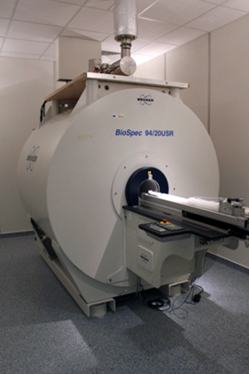
U28-E01. Bruker BioSpec 9.4T animal MRI system

U28-E01. Bruker BioSpec 9.4T animal MRI system


The Biospec 94/20 is a multipurpose research system for high-resolution magnetic resonance spectroscopy and imaging. The system is designed for preclinical and molecular MR imaging. State-of-the-art RF probes technology together with ultra high field USR magnets deliver high spatial resolution in vivo enabling researchers to come closer to the molecular and cellular level (molecular imaging). Thanks to its innovative modular concept, virtually any small animal MR imaging application in life science, biomedical and preclinical research can be conducted.
Specifications:
Magnetic field strength of 9.4T
20 cm bore diameter, suitable for mice and rats
Automated positioning system (AutoPac™)
High performance actively shielded
gradient system with integrated shims
RF coils for rats and mice

Read More

U22-E09. Metabolic cages to perform toxicity studies in rodents

Metabolic cages to perform toxicity studies in rodents

Read More

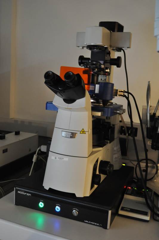
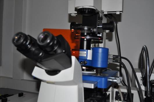
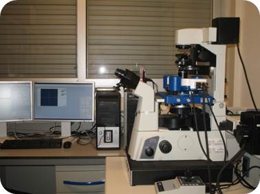
U13-E3. Atomic Force Microscopy

AFM: Atomic Force Microscopy JPK. AFMs can be used to measure the forces between the probe and the sample as a function of their mutual separation. This can be applied to perform force spectroscopy.


Read More

U12-E21. Fluorimeter Cary Eclipse (Varian)

U12-E21. Fluorimeter Cary Eclipse (Varian)

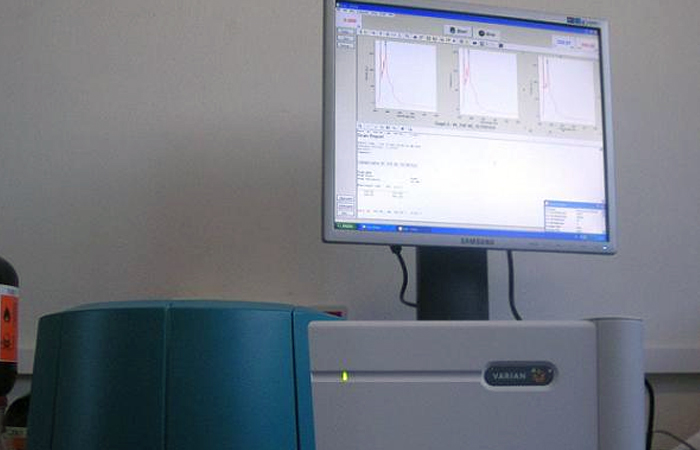
Fluorimeter Cary Eclipse (Varian) for fluorescence, phosphorescence, chemiluminiscence, bioluminiscence measurements.

U12-E21. Fluorimeter Cary Eclipse (Varian)
Read More

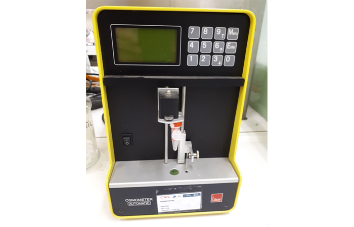
U12-E20. Osmometer Type 15 (Löser Messtechnik)

U12-E20. Osmometer Type 15 (Löser Messtechnik)

Description:

Instrument for the determination of osmolality via the measurement of freezing point

Applications:

Measurement of osmolality of biomedical samples

U12-E20. Osmometer Type 15 (Löser Messtechnik)
Read More

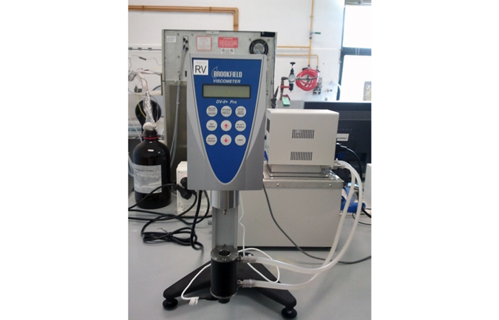
U12-E18. Viscosimeter RVDV-II+P (Brookfield)

U12-E18. Viscosimeter RVDV-II P (Brookfield)

Description:
Brookfield viscometer with a wide measurement range from 25 mPa.s to 64 MmPa.s. It is equipped with an accessory (Small Sample Adaptor SSA) for samples of small volume (7-13.5 mL).

Applications:
Viscosity measurements.

U12-E18. Viscosimeter RVDV-II P (Brookfield)
Read More

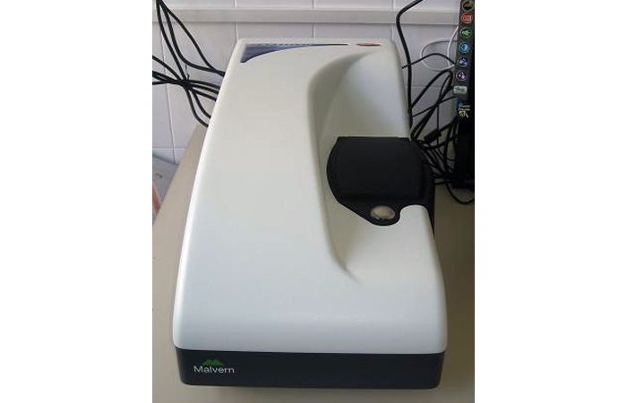
U12-E17. Zetasizer NanoZ (Malvern Instruments)

U12-E17. Zetasizer NanoZ (Malvern Instruments)

Description:
Instrument for the measurement of zeta potential, using the M3-PALS technology for the determination of electrophoretic mobility.  Temperature can be controled from 2 to 90 ˚C. It uses disposable capillary cells “Universal Dip Cell” for standard Zeta potential measurements in organic and aqueous solvents, and it can use the “High Concentration Cell“ for measurements at high concentrations

Applications:
Measurement of electrophoretic mobility and zeta potential in dispersions

U12-E17. Zetasizer NanoZ (Malvern Instruments)
Read More

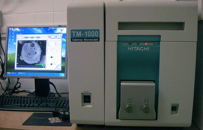
U12-E15. SEM Hitachi TM-1000 (Hitachi)

U12-E15. SEM Hitachi TM-1000 (Hitachi)

Description:
Tabletop Scanning Electron Microscope.

Applications:
Observations of the morphology of solid samples, such as polymeric and synthetic particles, meso/macroporous materials, textiles, etc., with high field depth (it allows focusing simultaneously on a wide portion of the sample) and relatively high resolution (in the nanometer range). Samples do not need to be electrical conductive, and therefore, coating with a very thin metal layer (e.g. Au, Pd) is not always required. The only sample requirement is the absence of volatile components, including water humidity

U12-E15. SEM Hitachi TM-1000 (Hitachi)
Read More

U20-E04. General laboratory equipment

General laboratory equipment

Read More