+34 620 10 75 37info@nanbiosis.com

Nanbiosis

In search of antimicrobials from natural bee products to coat implantable biomaterials, avoiding resistance.

The Microbial Adhesion research group-NANBIOSIS ICTS U16 Surface Characterization and Calorimetry Unit of the University of Extremadura (AM-UEX)-, belonging to the CIBER-BBN, led by Maria Luisa González, is searching in natural products, specifically in propolis, compounds with antimicrobial activity to help fight infections associated with biomaterials.

Medical devices have greatly improved healthcare. But biofilm-associated infections related to the use of these devices are a major clinical concern. Biofilms are understood as bacterial communities that adhere to the surface of the devices and are embedded in a polymeric matrix that they themselves produce. This supracellular social organization arises as a survival strategy in hostile environments, such as the human being itself, endowing the microorganisms embedded in it with resistance to mechanical clearance, the host’s immune response and antimicrobial agents. In this context, to prevent bacterial adhesion and the subsequent formation of biofilms, one of the prevention strategies is the coating of the biomaterial surfaces or the incorporation into the biomaterial itself of antimicrobial agents that can prevent their development. These type of infection are also aggravated by the multi-resistance of the microorganisms involved. For this reason, the AM-UEX group works in the search for natural products, with antimicrobial activity, that do not generate resistance, for their incorporation into new implantable biomaterials.

Bees are our allies, and their products can be a good source of available antimicrobials. Propolis is a glue for the hive and is a potentially useful food additive as it contains antioxidant and preservative properties. However, its application in other fields is limited, due to its strong flavor and low solubility. In addition, standardization is difficult because its chemical composition varies according to the flora of the environment. However, it’s common to all that they exhibit remarkable biological activities.

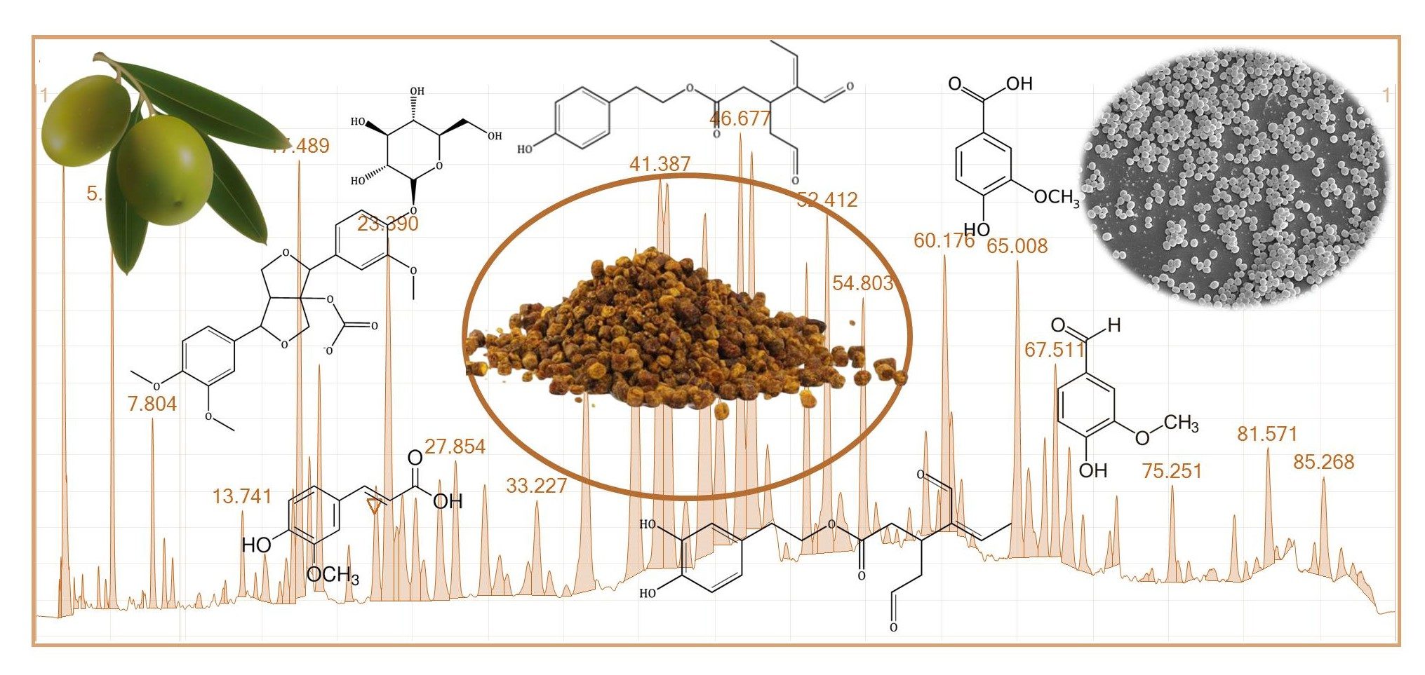
In a first study, the chemical composition of a Spanish propolis with a high antimicrobial capacity against bacterial strains closely related to infections associated with the formation of biofilms on biomaterials, Staphylococcus epidermidis, has been identified. The group has found in a novel Spanish ethanolic extract of propolis (SEEP) a high amount of polyphenols (205 ± 34 mg GAE / g), of which more than half correspond to the flavonoids group ( 127 ± 19 mg QE / g). The importance of this finding lies in the remarkable antioxidant and antimicrobial activities that have been attributed to this class of phenols. In addition, a more detailed analysis revealed the presence of compounds that are also present in olive oil such as vanillic acid, 1-Acetoxypinoresinol, p-HPEA-EA and 3,4-DHPEA-EDA, not previously detected in samples of propolis, which contribute to various health benefits. Other compounds found in relatively low amounts such as ferulic acid and quercetin also provide important therapeutic benefits. Regarding the antimicrobial properties of SEEP, a high sensitivity for S. epidermidis at low concentrations and a high inhibitory capacity at lower concentrations were found.

The antibacterial activity of propolis has been extensively studied, but its mechanism of action remains unclear. Research by our group has focused on measuring alterations in the physicochemical properties of the outermost surface layer of bacterial cells, both in gram-positive (S. epidermidis) and gram-negative (E. coli) cells, after incubation. with different concentrations of this antimicrobial agent. Propolis was found to induce substantial changes in bulk charge density, electrophoretic smoothness, and degree of hydrophobicity of the outermost surface layer of cells. Furthermore, observation by electron microscopy and determination of the release of cellular components carried out in NANBIOSIS Unit 16 of CIBER-BBN and UEX showed that propolis at sub-bactericidal concentrations already causes, at least locally, structural and morphological damage and/or disturbances in the cell wall. This research proposes that the mechanism of action of propolis against bacteria comes initially from the structural damage of the membrane / wall produced by the different constituents of propolis. It is a mechanism of action to which it can be difficult for bacteria to generate resistance, especially if different SEEP molecules work together synergistically.

Reference articles:

Fernández-Calderón, M. C., Navarro-Pérez, M. L., Blanco-Roca, M. T., Gómez-Navia, C., Pérez-Giraldo, C., and Vadillo-Rodríguez, V. (2020). Chemical Profile and Antibacterial Activity of a Novel Spanish Propolis with New Polyphenols also Found in Olive Oil and High Amounts of Flavonoids. Molecules 25, 3318. [DOI]

Vadillo-Rodríguez V, Cavagnola MA, Pérez-Giraldo, Fernández-Calderón MC. (2021) A physico-chemical study of the interaction of ethanolic extracts of propolis with bacterial cells. Colloids Surf B Biointerfaces 200, 111571. [DOI]
Read More

Extracellular vesicles as vehicles for therapeutic enzymes in lysosomal deposition diseases

Researchers of two CIBER-BBN units of the ICTS (Singular Scientific and Technical Infrastructures) NANBIOSIS, led by Ibane Abasolo the U20 located in the VHIR and the U6 in ICMAB-CSIC have participated in a study to improve the treatment of lysosomal disorders.

In recent years, enzyme replacement therapies (ERTs) based on the systemic administration of a functional version of the defective enzyme have gained clinical relevance as a treatment for lysosomal storage disorders (LSD). However, the systemic administration of these recombinant enzymes has negative aspects, such as their low stability and inadequate distribution to the affected organs that result in a limited efficacy of ERT. In this context, the Vall d’Hebron Research Institute (VHIR) has led a study that has analysed the use of extracellular vesicles as vehicles for therapeutic enzymes in this type of lysosomal storage disorders. This work, in which the CIBBIM-Nanomedicine, Drug Delivery and Targeting, led by Dr. Ibane Abasolo and investigator Guillem Pintos, and Neurovascular Diseases, led by Dr. Anna Rosell, of the VHIR have participated, has been published in the Journal of Extracellular Vesicles.

Lysosomal storage disorders are rare congenital diseases caused by the lack or malfunction of proteins involved in lysosomal biogenesis and activity. In the absence of activity of these proteins, lysosomes accumulate waste molecules inside. Although there are more than seventy types of LSD – among which are the Gaucher, Fabry, Pompe and Sanfilippo syndromes – all of them share the abnormal accumulation of molecules such as glycoproteins, glycosaminoglycans and sphingolipids, something that in the majority of the cases generates serious clinical manifestations.

The symptoms that the patient may experience depend on the specific disorder they develop, but in general LSDs results in a systemic disease that can affect multiple organs, including the central nervous system, liver, kidneys, heart and musculoskeletal system”, says Dr. Ibane Abasolo, principal investigator of the research group in CIBBIM-Nanomedicine, Drug Delivery and Targeting of the VHIR and author of the study. All LSDs are rare diseases and their low incidence makes it difficult to develop new therapies and evaluate them in clinical trials. For all these reasons, the VHIR has wanted to participate in this work, which has analysed and tested the viability of a new therapy for this type of lysosomal storage disorders. Specifically, for Fabry and Sanfilippo A diseases, two of the LSDs with the highest prevalence.

The study has obtained extracellular vesicles loaded with therapeutic enzymes directly from cells that are used for the production of recombinant protein. Extracellular vesicles have been shown to function as highly efficient protein delivery platforms, also in the brain, an organ that is not naturally accessed by systematically administered proteins. Dr. Abasolo considers that “the results obtained in this work will lay the foundations for the implementation of replacement therapy driven by extracellular vesicles in different LSDs, and will open the way for a possible treatment strategy in disorders with central nervous system involvement”.

The following centres have also participated in this work: Institute of Biotechnology and Biomedicine of the Autonomous University of Barcelona (IBB-UAB), Centre for Biomedical Research in Network of Bioengineering, Biomaterials and Nanomedicine (CIBER-BBN), Institute of Bioengineering of Catalonia (IBEC), Institute of Advanced Chemistry of Catalonia (IQAC-CSIC), and Centre for Biomedical Research in Network on Liver and Digestive Diseases (CIBEREHD).

Read More

The relevance of biomedical signal processing in the understanding of biological systems

Within the framework of NeoCom2021 Jesús Lázaro, researcher of BSICoS group and NANBIOSIS U27 High Performance Computing form CIBER-BBN and I3A-UZ will explain how biomedical signal processing can be used to improve the current understanding of the functioning of biological systems, conditions related to the cardiovascular, respiratory, and autonomic nervous systems, as well as their interactions.

Prof. Lázaro will review the progress of the WECARMON European Project whose objective is the development of a system for long-term monitoring (months / years) of patients with cardiorespiratory diseases.

NEOCOM: As every year, the Territorial Demarcation of the COIT in Aragon and the Association of Telecommunications Engineers of Aragon collaborate with the Association of Telecommunications Students of the University of Zaragoza (AATUZ) in the organization of the NEOcom conferences that bring ICT companies closer to the university field. All talks are broadcasted on live on the AATUZ YouTube channel (without registration)

Jesús Lázaro and Wecarmon Project

Related news:

NANBIOSIS U27 researchers working in an App for the early diagnosis of covid-19 through mobile phones

Wearable Armband Device for Daily Life Electrocardiogram Monitoring

Read More

Development of a protein-only drug delivery system for sustained release

The Nanotoxicology Unit of CIBER-BBN ICTS NANBIOSIS (u18-nanotoxicology-unit), led by Ramon Mangues at the Research Institute of the Hospital de Sant Pau and the NABIOSIS (nanbiosis.es)Protein Production Platform (u1-protein-production-platform-ppp) led by Antonio Villaverde and Neus Ferrer Miralles of the Institute of Biotechnology and Biomedicine at the Autonomous University of Barcelona, have participated in the development of a novel drug delivery system able to achieve sustained release of proteins with intrinsic antitumor activity. This delivery system consists on inclusion bacterial bodies formed by recombinant fusion proteins that precipitate while being expressed in bacteria, acquiring an amyloid structure, but remaining functional. Thus, these amyloids are able to release protein monomers that generate soluble nanoparticles that selectively internalize within target cancer cells because of the incorporation ion the protein nanoparticles of a specific ligand that interacts with a surface receptor expressed in target cancer cells.

On this basis, we have subcutaneously administered inclusion bodies containing cytotoxic nanoparticles that incorporate the Pseudomonas aeruginosa Exotoxin (PE24), to demonstrate their capacity of sustained release since they reach cancer tissues through the blood to selectively killing target colorectal (CRC) cancer cells. This cancer specific targeting occurs because the released protein is functionalized with the peptidic ligand T22 for the CXCR4 receptor (overexpress in CRC cells). In addition, we have evaluated their anticancer effect in the different localization where metastatic foci growth in a colorectal cancer (CRC) model. The administration, in these models of 500 micrograms of T22-PE24 amyloids, induces a potent inhibition of primary tumor growth and a dramatic reduction, both in number and size, of the metastases in lymph nodes, liver, lung and peritoneum, an effect that is achieved, in the absence of systemic toxicity.

One of the main applications of these functional amyloid structures could be their use by subcutaneously injectable drug depots that could release the active protein drug at sustained levels, during a long time (weeks or months). This may change the way that current protein-based drugs (e.g. antibodies targeting specific receptors) are administered, since this approach will allow to dosage the drug only once every 2-3 weeks or a month, avoiding the current intravenous injection schedule, which is 2-3 times a week. When applied to targeted drugs, as the one here described, which demonstrate high efficacy with low adverse effects, could establish a novel approach to treat cancer patients by visits of sanitary personnel at their home, avoiding the need of their hospitalization, which is required when receiving intravenous injections; and therefore, dramatically reducing the cost of patient treatment for the health system. 

Reference:

Céspedes MV, Cano-Garrido O, Álamo P, Sala R, Gallardo A, Serna N, Falgàs A, Voltà-Durán E, Casanova I, Sánchez-Chardi A, López-Laguna H, Sánchez-García L, Sánchez JM, Unzueta U, Vázquez E, Mangues R, Villaverde A. Engineering Secretory Amyloids for Remote and Highly Selective Destruction of Metastatic Foci. Advanced materials. 2020.  https://doi.org/10.1002/adma.201907348

Read More

A new generation of devices for the rapid, cheap and easy diagnosis of candidemia.

Candidemia is an infection caused by fungi of the genus Candida that is associated with high complication rates with a mortality that can reach 40%. A group of researchers have managed to develop a new material that allows for the rapid detection of candidaemia, with a sensitivity that exceeds 90% and a specificity greater than 95%.

Researchers from the CIBER-BBN (Center for Biomedical Research for Bioengineering, Biomaterials and Nanomedicine). the Polytechnic University of Valencia (UPV) and the La Fe Health Research Institute (IIS La Fe), with the participation of Unit 26 of NANBIOSIS NMR Biomedical Applications II, in collaboration with the Valencian startup MATCH Biosystems have developed a new generation of in vitro diagnostic devices (IVD), capable of improving current times, with high reliability and easy handling.

“The characterization of the anchored molecules as well as the final sensors were characterized using NMR equipment from NANBIOSIS unit 26”, explains Ramón Martínez Máñez, Scientific Director of NANBIOSIS Unit 26 of NMR Biomedical Applications of the University of Valencia and the CIBER-BBN

The group of researchers, led by Ramón Martínez Máñez, professor of Inorganic Chemistry at the Polytechnic University of Valencia and scientific director of the CIBER-BBN; and Javier Pemán, head of the Mycology Unit of the Microbiology Service of the Hospital Universitari i Politècnic La Fe, and head of the Severe Infection Research Group at IIS La Fe, decided to create MATCH biosystems in June 2020 with the objective of marketing in the future “the new generation of IVD devices capable of obtaining results for the diagnosis of candidemia in less than an hour in a reliable, easy way and without the need to go through a laboratory”, explains Adrián Teruel, CEO of the biomedical startup, pharmacist, doctor in nanomedicine.

Currently, the results of candedimia infection are obtained after carrying out cultures of the biological fluid in the laboratory, which can take between three and four days. With the new material and method patented by the researches’ institutions and developed by MATCH biosystems, the diagnostic time is drastically reduced.

“The tests are carried out very quickly, easily and without the need to enter a laboratory, which also reduces the equipment and personnel necessary to carry them out, with the consequent reduction in the expense that this implies for health public, in addition to allowing speed in making medical decisions that can save lives “, details Adrián Teruel.

The material used by MATCH biosystems is composed of dye-loaded porous supports and single strands of DNA, which act as molecular gates. The infection is detected when the single strands of DNA recognize the genetic material of the pathogen in the patient sample, at which point the molecular gates are opened and the dye is released, allowing the diagnosis to be carried out. The first sensors have already been developed and clinical tests have been carried out with real samples of patients, at the Hospital de La Fe.

MATCH biosystems is currently working on developing the complete kit that it expects to reach the market in a maximum of four years, in parallel with obtaining the relevant regulations and certifications.

The company has now focused on infections caused by candidemia, “but the field of application of the new devices also includes, in addition to the clinical, the veterinarian and the agri-food industry” says Teruel.

Related news

Read More

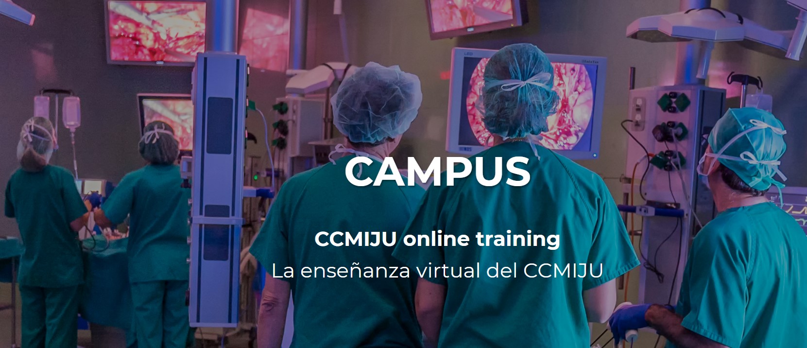
CAMPUS JUMISC online training

The Jesús Usón Minimally Invasive Surgery Center, CCMIJU, partner of NANBIOSIS, offer high quality training based on itslearning philosophy perfectly adapted to new teaching methodologies and to a more dynamic and interconnected world.

The CCMIJU Campus responds to the new modalities of online training and education. Based on their interests and objectives, students choose the course to suit their level and learning pace, being able to interact with expert tutors through social networks. Emerging as a complement to face-to-face activity, the CCMIJU Campus offers high quality training adapted to new methodologies, and to a more dynamic and intercommunicated world.

The only training modality provides a self-paced learning for those who need to start completing the targets at any time and can arrange their schedule to satisfy individual needs. All lectures, videos and needed materials are provided via this online platform.

The face-to-face courses offer a pyramid training system with two training modalities: Set courses (Scheduled activities monitored by trainers of different specialties) and Under-request courses (Training courses on minimally invasive surgery techniques, addressed to medicine professionals to improve their skills in the use of new techniques, devices and equipment)

Further information: https://www.campusccmiju.com/

Read More

Modulation of intercolumnar synchronization by endogenous electric fields in cerebral cortex with neuroprobes by NANBIOSIS unit 8

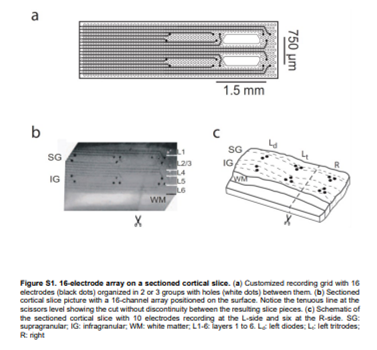
The collaboration of NANBIOSIS U8 Micro–Nano Technology Unit of the CIBER in Bioengineering, Biomaterials & Nanomedicine (CIBER-BBN) at the IMB-CNM in the research carried out by scientist of Institut d’Investigacions Biomèdiques August Pi i Sunyer (IDIBAPS), Université Paris-Saclay, Centre National de la Recherche Scientifique (CNRS) and ICREA has bien acknowledged in the publication of the results by Science Advances. Rosa Villa and Xavi Illa have been in charge of the fabrication of the probes used, more specifically, neuroprobes were designed and manufactured with 16 Ti / Au microelectrodes (20 / 200nm) on flexible polyimide substrates with open areas to improve neuronal tissue viability according to specifications.

Abstract

Neurons synaptically interacting in a conductive medium generate extracellular endogenous electric fields (EFs) that reciprocally affect membrane potential. Exogenous EFs modulate neuronal activity, and their clinical applications are being profusely explored. However, whether endogenous EFs contribute to network synchronization remains unclear. We analyzed spontaneously generated slow-wave activity in the cerebral cortex network in vitro, which allowed us to distinguish synaptic from nonsynaptic mechanisms of activity propagation and synchronization. Slow oscillations generated EFs that propagated independently of synaptic transmission. We demonstrate that cortical oscillations modulate spontaneous rhythmic activity of neighboring synaptically disconnected cortical columns if layers are aligned. We provide experimental evidence that these EF-mediated effects are compatible with electric dipoles. With a model of interacting dipoles, we reproduce the experimental measurements and predict that endogenous EF–mediated synchronizing effects should be relevant in the brain. Thus, experiments and models suggest that electric-dipole interactions contribute to synchronization of neighboring cortical columns.

Article of refrence:

Modulation of intercolumnar synchronization by endogenous electric fields in cerebral cortex. Beatriz Rebollo, Bartosz Telenczuk,  Alvaro Navarro-Guzman,  Alain Destexhe and Maria V. Sanchez-Vives Science Advances  03 Mar 2021: Vol. 7, no. 10, eabc7772 DOI: 10.1126/sciadv.abc7772

Read More

Simo Schwartz: new General Director of the Bank of Blood and Tissues of Catalonia.

Simó Schwartz Jr., Scientific Director of NANBIOSIS Unit 20 In Vivo Experimental Platform has been appointed as the new general director of the Bank of Blood and Tissues of Catalonia, replacing Enric Argelagués, who is leaving his post after 17 years as director. Argelagués was the promoter of the unification of blood banks that began in the eighties and culminated in the current Bank of Blood and Tissues of Catalonia.

Simó Schwartz now assumes the executive management of the organization, which currently has more than 800 workers and its mission is to ensure that all people in Catalonia have the blood and tissues necessary for their treatment at their disposal, promoting proper use. It is a reference center in diagnostic immunology and in the development of advanced therapies, it is present in the main hospitals in Catalonia and is part of the national and international organizations related to donation, transfusion and treatments with biological components.

Read More

Reversing pulmonary fibrosis has been achieved in murine models with peptide-loaded nanoparticles

A new study with participation of NANBIOSIS Unit 10 Drug Formulation unit of CIBER-BBN and UPV/EHU, has succeeded in preventing and reversing pulmonary fibrosis in experimental models with PLGA / PEI nanoparticles loaded with the GSE4 peptide. These results are the first step to study its possible antifibrotic effect in patients with idiopathic pulmonary fibrosis and other progressive fibrosing diffuse interstitial lung diseases that present altered telomere function.

NANBIOSIS unit 10 has been responsible for the design and manufacture of biodegradable nanoparticles loaded with the peptide.

The work is led by Rosario Perona and Leandro Sastre, researchers of CIBERER at the Biomedical Research Institute (CSIC/UAM), and Maria Molina-Molina, researcher at CIBERES and the Department of Pulmonology at the Bellvitge University Hospital. In addition, Guillermo Güenechea, from CIBERER at CIEMAT-IIS / FJD, and José Luis Pedraz, from CIBER-BBN and the University of the Basque Country have collaborated.

Idiopathic pulmonary fibrosis is a fatal fibrotic disease associated with aging with a median survival of 2 to 5 years. The disease usually appears around the age of 60, but there are familiar forms that can manifest at younger ages. Fibrosis appears as an aberrant healing response after alveolar damage and is characterized by the appearance of abnormal lung scarring, in which the restoration of epithelial integrity and tissue function is compromised. The mechanisms responsible for defective repair and regeneration are not well understood. However, recent studies suggest a possible role of accelerated aging and telomere shortening in the onset of the disease, mainly in familial forms.

The GSE4 peptide, of 11 amino acids, corresponds to an internal sequence of dyskerin, a protein that is part of the telomerase complex. GSE4 has protective effects on the cells of patients with the rare diseases dyskeratosis congenita and ataxia-telangiectasia, as it increases telomerase activity and cell growth and reduces DNA damage, oxidative stress and cell senescence.

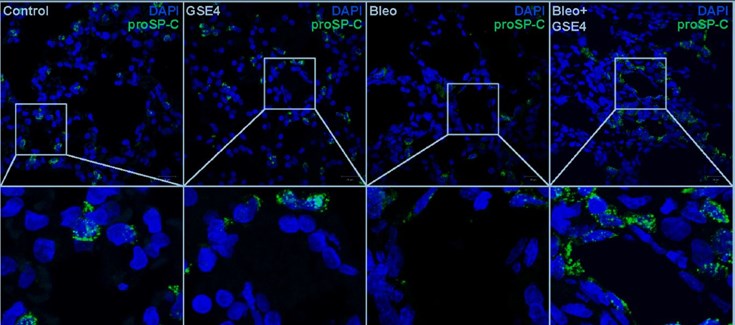
The authors of the study, published in The FASEB Journal, used rat lung alveolar cells treated with a profibrotic agent, bleomycin, as a model. Expression of the GSE4 peptide or treatment with GSE4-PLGA / PEI nanoparticles causes a reversal of the inflammatory and fibrotic phenotype. There is an increase in telomerase activity, a decrease in DNA damage, and a decrease in inflammation and cell death. Furthermore, these cells show an inhibition in fibrosis.

In a rat model with bleomycin-induced pulmonary fibrosis, treatment with GSE4-PLGA / PEI nanoparticles has also increased telomerase activity and decreased DNA damage in alveolar cells. In both preventive and therapeutic protocols, GSE4-PLGA / PEI nanoparticles have prevented and attenuated lung damage and inhibited fibrosis. Histological analysis of the lungs of rats treated with bleomycin and GSE4-PLGA / PEI nanoparticles shows less fibrosis and inflammation and greater regeneration of the alveolar tissue, indicating the therapeutic efficacy of GSE4-loaded nanoparticles in this experimental pulmonary fibrosis model. .

This study supports the possibility of further investigating a potential curative treatment for patients with pulmonary fibrosis.

Reference article:

Pintado-Berninches, L., Montes-Worboys, A., Manguan-García, C., et al. “GSE4-loaded nanoparticles a potential therapy for lung fibrosis that enhances pneumocyte growth, reduces apoptosis and DNA damage.” The FASEB Journal. 2021; 35: e21422. DOI: 10.1096/fj.202001160RR

(*) Explanation of the figure: Treatment with the profibrotic agent bleomycin decreases the number of cells that express the alveolar marker proSP-C (Bleo). Inoculation of GSE4-loaded nanoparticles into bleomycin-treated rats increases the number of cells expressing the marker, indicating alveolar regeneration.

Read More

Graphene sensors read low-frequency neural waves associated with distinct brain states

Xavier Illa, Anton Guimrea y Eduard Masvidal, researchers of the CIBER-BBN group GAB Lab at IMB-CNM led by Rosa Villa, are coauthors of a study recently published in “Nature Communication”, in which it is demonstrated that graphene-based active sensor arrays are a mature technology for large-scale application in wide frequency band neural sensing interfaces. NANBIOSIS U8 Micro– Nano Technology Unit has been used in the development of the research.

This research has been carruied out within the framework of the European Project “Graphene Flagship. The scientists have developed a sensor based on CVD graphene that detects brain signals in a wide frequency band, from extremely low frequencies to high frequency oscillations. The sensor is biocompatible and could be used to measure and predict brain states. Furthermore, the graphene sensors could be used in chronic implants due to their high stability in the brain.

Further information: News by the Graphene Flagship website.

Article of reference:

Garcia-Cortadella R, Schwesig G, Jeschke C, Illa X, Gray AL, Savage S, Stamatidou E, Schiessl I, Masvidal-Codina E, Kostarelos K, Guimerà-Brunet A, Sirota A, Garrido JA. Graphene active sensor arrays for long-term and wireless mapping of wide frequency band epicortical brain activity.  Nat Commun 12, 211 (2021). https://doi.org/10.1038/s41467-020-20546-w

Read More