Specifications:
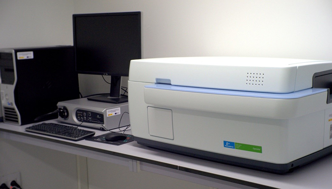
Spinning disc confocal technology
10x, 20x and 40x long working distance objectives
20x high NA objectives
Temperature control and CO2 for cultured cells
à 400W Xenon based illumination
Fully automated image acquisition from 24,96 and 384 well plates
Harmony: integrated acquisition and analysis software
Phenologic – machine learning-based phenotype recognition software
Columbus Image Data Storage and Analysis System

Specifications:
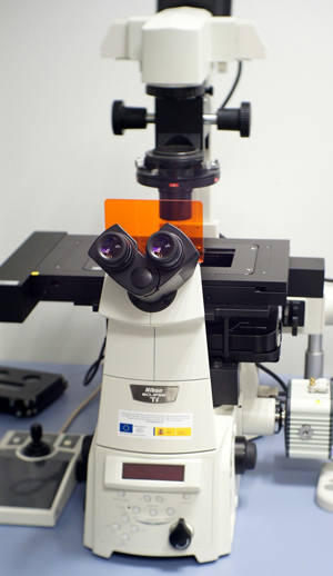
Inverted Nikon Ti Eclipse fully motorised microscope
Hamamatsu Orca R2 monochrome 1.3 MP CCD camera
Okolab H101 stage-top incubator with temperature, humidity and CO2 control
Objective lenses:
Nikon Intensilight 130w Mercury Lamp
Filter sets for DAPI, GFP, TRITC
Nikon PFS (Perfect Focus System) for improved stability

Specifications:
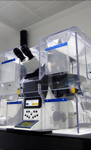
Inverted Nikon Ti Eclipse fully motorised microscope
100x Plan Apo Oil immersion Objective 1.49 NA
TIRF optical system (used to improve contrast with N-STORM samples)
Motorised TIRF mirror
Multipass TIRF dichroic filter (allows high-speed multicolour TIRF)
Andor iXon3 897 EM-CCD camera
Microscope enclosure with temperature, humidity and CO2 control
High precision motorized stage.
Nikon PFS (Perfect Focus System) for improved stability
Offline workstation with additional processing module
Objective lenses:
4 Lasers in the visible range:

Specifications:
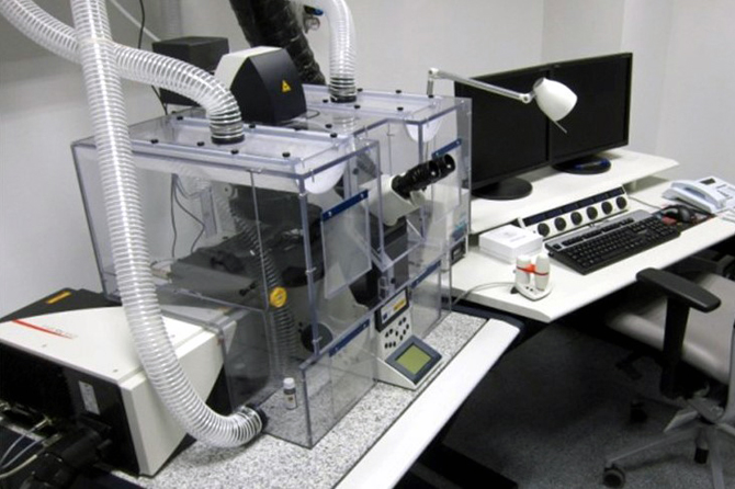
Inverted Leica DMI 6000 fully motorised microscope
Confocal detectors: 2 x HyD, 3 x PMT + 1 transmitted light detector
Tandem Scanner – HQ conventional scanner and high-speed Resonant scanner (28 frames/sec @ 512 x 512)
AOBS (Acousto-Optical beam splitter) – brighter and more flexible than con-ventional beam splitters
Optical zoom up to 64x (maximum resolution is dependent on the objective used)
Choice of objectives:
9 Laser lines in the visible range:
High Performance Mai-Tai titanium-sapphire IR femtosecond pulse laser for multi-photon imaging, tunable from 690-1040nm (maximum power 800nm∼ 2.9W)
Microscope enclosure with temperature, humidity and CO2 control
High precision Super Z motorized stage with multi-point and multi-tile soft-ware control. 3nm resolution galvometric z-axis control

Specifications:
Inverted Leica DMI 6000 fully motorised microscope
Confocal detectors: 2 x HyD, 3 x PMT + 1 transmitted light detector
Tandem Scanner – HQ conventional scanner and high-speed Resonant scanner (28 frames/sec @ 512 x 512)
AOBS (Acousto-Optical beam splitter) – brighter and more flexible than con-ventional beam splitters
Optical zoom up to 64x (maximum resolution is dependent on the objective used)
Choice of objectives:
9 Laser lines in the visible range:
Microscope enclosure with temperature, humidity and CO2 control
High precision Super Z motorized stage with multi-point and multi-tile soft-ware control. 3nm resolution galvometric z-axis control
Specifications:
Working temperature 4 – 60 °C (at an ambient temperature range between 18 – 25 °C).
Peltier controlled heating/cooling.
Maintain relative humidity at 100 %.
Ultrasonic controlled humidification.
Linux operating system.
Touch screen control and set-up. Mouse and foot pedal controls also available.
Small sample volumes can be applied manually with a pipette through a small side port on the left and the right side of the climate chamber.
Both application time and wait time (between application and blotting) are software controlled and can be set in the user interface.
Precisely timed control of multiple sample applications, blotting actions, and vitrification enables time resolved analysis of interactions among separately applied components.
Excess fluid is removed from the grid by (repeated) blotting with filter paper on rotating foam pads.
Number of blotting actions (max. 16 times for one grid) and duration of blotting are software controlled and can be set in the user interface.
Longitudinal grid positioning (‘blot offset’) and wait time between blotting and vitrification (‘drain time’) are user definable.
Automated shutter control allows smooth, instant injection of the sample grid into the coolant (liquid ethane or propane). A lift for the container brings the coolant close to the shutter to ensure optimal vitrification.
Synchronous lowering of the coolant container and the grid holder keeps the grid submerged in the coolant and minimizes the risk of contamination prior to transfer ring the sample into a storage box or cryo holder.
Coolant container including an integrated anti-contamination ring.
Specifications:
Reproducible operations.
Excellent specimen visibility with internal LED illumination.
Working range from -140 °C to +70 °C.
“Deep Freeze” allows sample transfer at temperatures below -140 °C.
Transfer function “TF” excludes humidity and oxygen.
Fast UV polymerization with LED UV lamp.
Any choice of substitution system, e.g.:
Mouse controlled color screen.
Memory stick to transfer programs.
Log file download on memory stick.
35 liters Dewar for liquid nitrogen.
Liquid nitrogen filling from outside specimen chamber, with a consumption enough for 5 day protocols.
Stainless steel working platform.
The Leica EM FSP (“Freeze Substitution Processor”) is an automatic reagent handling system. Mounted on the Leica EM AFS2, it dispenses reagents for both FS and PLT. It automatically dilutes FS media and resins from 100 % reagent containers. An integrated LED UV lamp allows immediate polymerization of the samples.
Specifications:
• Automatic freezing of samples with one button operation.
• Extremely high cooling rates.
• Perfect freezing of samples up to 6 mm diameter.
• No cryoprotectants required.
• Rapid transfer loading device for CLEM (‘Correlative Light and Electron Microscopy’) work.
• Fast, ergonomic sample loading.
• Express sample handling for fast freezing.
• Graphical data display of actual temperature, time and pressure for each run.
• Integrated work station.
• Integrated stereomicroscope and adjustable LED illumination.
• Universal and application specific sample carriers.
• Integrated Dewar with drain outlet.
• Automatic bake-out cycle.
• Thermally insulated process chamber.
• Integrated touch control screen with prompt window.
• Freezing process cover for ultimate safety.
• USB interface for data storage and transfer.
• Integrated air compressor.
Specifications:
• Using the touchscreen control, three different cryo-modes are available:
– Standard.
– High gas flow, increased gaseous nitrogen flow reduces ice contamination below -140 °C.
– Wet sectioning, to set a temperature difference of up to 130 °C between knife (-40 °C) and specimen (-170 °C), which is useful for, e.g., DMSO applications.
• Leica EM CRION ionizer with electrostatic charge and discharge functions controlled by foot switches greatly improves the method of cryosection collection.
• The attachable micromanipulator provides precise positioning of the grid to make section collection easier than ever before.
• Multiple lighting angles using the patented LED chamber illumination provide excellent visibility for section manipulation and pick up.
• Contact-free, through-the-wall specimen arm makes the instrument highly stable for chatter-free cryosectioning.
• Heated chamber walls prevent icing over a prolonged working time and enhance user comfort.
• Especially for frozen hydrated sectioning or the Tokuyasu method, the Leica EM CRION with electrostatic discharge and charge mode used with the micromanipulator provides outstanding cryo sectioning performance.
