+34 620 10 75 37info@nanbiosis.com

Nanbiosis

Institutional impulse to accelerate 3D printing projects

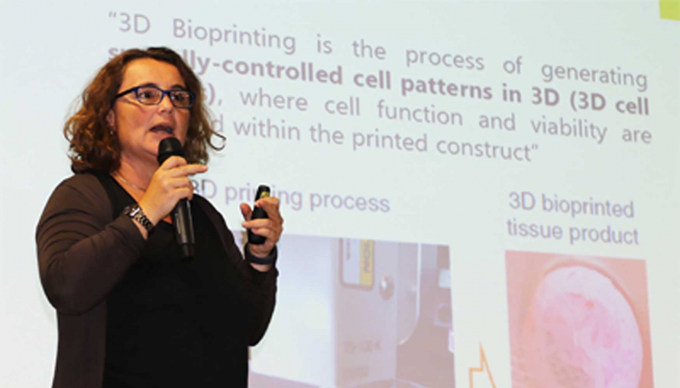
Elisabeth Engel,  Scientific Director of Unit 5 of NANBIOSIS, U5. Rapid Prototyping Unit, will led the research project, QuirofAM, to produce biodegradable and bioactive scaffolds printed in 3D as osteochondral and maxillofacial substitutes.

This is one of the projects to be promoted by LLAVOR 3D Community, an association of Catalan entities, created last month, which includes the IBEC (housing units 5 and 7 of NANBIOSIS). LLAVOR 3D Community aims to accelerate the development and adoption of additive manufacturing and 3D printing technologies by the industry.

Within the projects of the LLAVOR 3D Community, new software tools, new materials, more efficient and versatile production processes, new post processes and surface treatments as well as new 3D printing applications will be developed, contributing to the creation of an R & D ecosystem in additive manufacturing technologies reinforcing the position of Catalonia as an international benchmark.
Read More

Intestinal histopathological changes in a porcine model of pneumoperitoneum-induced intra-abdominal hypertension

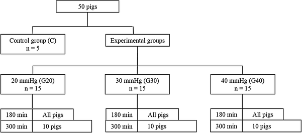
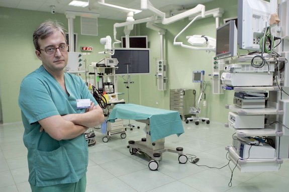
Francisco Miguel Sánchez Margallo, Scientific Director of  JUMISC, is co-author of the publication “Intestinal histopathological changes in a porcine model of pneumoperitoneum-induced intra-abdominal hypertension” by Surgical Endoscopy.

The study was performed with the participation of NANBIOSIS Units: U21-Experimental operating rooms, and U22-Animal housing. and showed that the  evolution of intestinal injuries from pneumoperitoneum-induced IAH depends on the degree of IAP, these damages may be associated with decreases in APP and pHi, and increases in Lc

For further information: click here

Read More

Why research in micro-electronics? Neural interfaces, interaction between the nervous and the artificial system

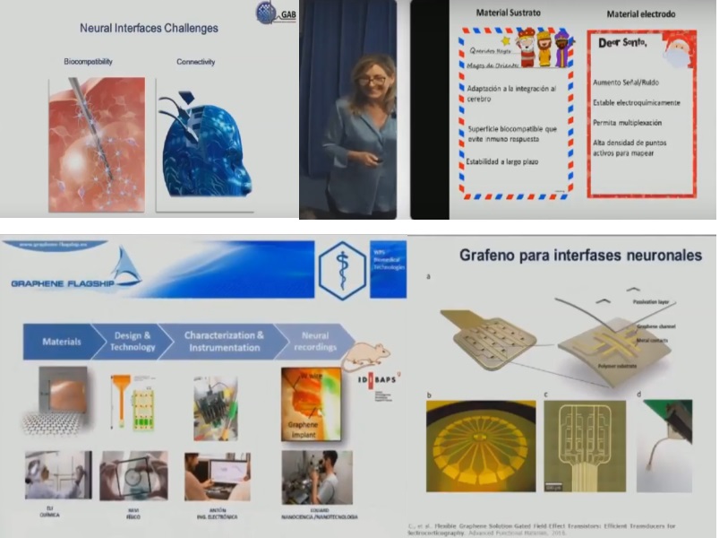
Dra. Rosa Villa,  Scientific Director of NANBIOSIS U8. Micro – Nano Technology Unit, explained last June, 11 at the Residence of Researchers of Barcelona the great scientific challenges in finding tools that allow a good interaction between the nervous and the artificial system.

This talk is part of the series of conferences organized by the CNM with the theme “Why research in micro-electronics?” Dr. Manuel Lozano, Director of the CNM, introduced the talk explaining that with microelectronic technology scientists  can capture the signals that occur in the nerves and in the brain and presented Rosa Villa  as a doctor  with a PhD in cochlear implants (electronic medical devices that perform the work of damaged parts of the inner ear (cochlea) to provide sound signals to the brain). Dr. Villa now directs the group of biomedical applications of the CNM that currently uses nano technologies in their research. Her training in medicine and microelectronics has allowed her to tackle electronic-based projects with biomedical application. Her lines of research are focused, nowadays, on neural interfaces and on organ-on-chip technology, (a multi-channel 3-D microfluidic cell culture chip that simulates the activities, mechanics and physiological response of the entire organs and organ systems, the group of Dr. Villa works mainly the liver, very useful for the test of drug patho-toxicity).

In this talk, Rosa Villa explains her second line research of neural interfaces, the development of suitable interfaces between the biological systems and electronic devices and how they study the improvement of the necessary technologies to restore the motor skills or to know how the brain works applying microelectronic techniques.  These investigations run into the main problems of biocompatibility and conectivity and Rosa Villa shows us her letter to Santa Claus to solve them and how graphene is being of great help.

You can see the whole conference clicking here

The video includes a funny class by Eli Prats to produce graphene at home.

Read More

Interpretation of motion analysis of laparoscopic instruments based on principal component analysis in box trainer settings

Francisco Miguel Sánchez Margallo, Scientific Director of  CCMIJU and NANBIOSIS U21. Experimental operating rooms, is co-author of the publication “Interpretation of motion analysis of laparoscopic instruments based on principal component analysis in box trainer settings” published by Surgical Endoscopy

The study results show that three new HSMAPs per hand were defined for PG and PC tasks, and two per hand for KS task. PG presented validity for HSMAPs related to insecurity and economy of space. PC showed validity for HSMAPs related to cutting efficacy, peripheral unawareness, and confidence. Finally, KS presented validity for HSMAPs related with economy of space and knotting securitya. Thus, PCA-defined HSMAPs can be used for technical skills’ assessment. Construct validation and expert knowledge can be combined to infer how competences are acquired in box trainer tasks. These findings can be exploited to provide residents with meaningful feedback on performance. Future works will compare the new HSMAPs with valid scoring systems such as GOALS.

The study has been develloped with surgical facilities of high technology that allow in vivo efficacy assays of drugs, nanomedicines, biomaterials and others, performed at unit 21 of NANBIOSIS.

Article of reference: Oropesa, I., Escamirosa, F.P., Sánchez-Margallo, J.A. et al. Surg Endosc (2018) 32: 3096. https://doi.org/10.1007/s00464-018-6022-6

Read More

NANBIOSIS U2 incorporates a new Nitrogen tank to store cells

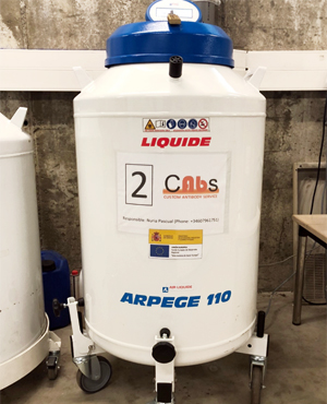
NANBIOSIS U2. Custom Antibody Service (CAbS), is incorporating new equipment as a result of its participation in the project FICTS1420-27, selected by the MINECO for co-financing by the FEDER Program in ICTS 2014-2020.

Recently the purchase and installation and set-up of a new nitrogen tank to store cells has taken place. The storage of cell lines of monoclonal antibody-producing hybridomas is a process in which the cells are conserved when they are cooled to temperatures typically at -196 ° C in liquid Nitrogen tanks. With temperatures below -130 ° C the activity Biological ceases and storage at these temperatures is vital to keep the material unaltered. The increasing activity of the NANBIOSIS U2 the makes necesary to incorporate a new tank of liquid Nitrogen for the storage of new hybridoma lines.

For further information:


Liquid nitrogen tank
Read More

Graphene will be manufactured with greater economic and biomedical benefits at NANBIOSIS unit 8

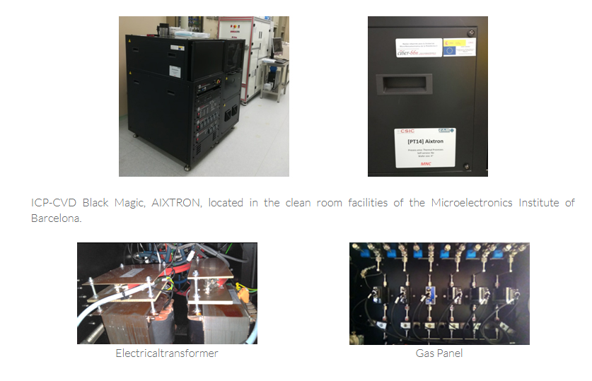
The unit 8 of NANBIOSIS U8-Micro, nanotechnology, will offer HIGH-QUALITY GRAPHENE GROWN ON ONE-layer or multi-layer CU SHEETS, as well as graphene transferred to different rigid and flexible parylene substrates while preserving their valuable initial properties

This unit has recently  upgraded  the Aixtron Black Magic Pro 4-inch System for the growth of graphene. It has also been purchased, installed and set-up by the unit parylene deposition device on flexible substrates. With this acquisition the unit will be able to offer high quality graphene grown in monolayer or multilayer CU sheets, as well as graphene transferred to different rigid and flexible parylene substrates while preserving their valuable initial properties.

ICTS NANBIOSIS currently has a CVD equipment from the Aixtron house (model Aixtron BM 4 “) that is specifically designed for the growth of nanotubes and carbon nanofibers on defined areas, which in turn are compatible with the growth of graphene, However, due to differences in the temperature conditions of the growth of these different materials, this machine requires an upgrade to Aixtron BM Pro 4 “, which is the one contemplated here. This update has been previously made with success in other equipment installed in the United States, such as the Texas Instruments company.

The growth of graphene can be done on different substrates both rigid (pyrex, silicon, silicon carbides) and flexible polymers such as SU8, polymides etc. Currently in the ICTS NANBIOSIS are optimized the processes for the manufacture of devices in rigid and flexible substrates of SU8 and polymidas and now with this new acquisition will include the manufacture with materials such as parylene that will provide greater profitability in the processes as well as greater benefits biomedical and

This upgrade has been confinanced by the European Regional Development Fund (ERDF) through the Plurirregional Operational Program of Spain (POPE)2014-2020

European Regional Development Fund

Read More

Update and improvement of unit 4 of NANBIOSIS

NANBIOSIS U4-Biodeposition and biosensing, led by Prof Laura Lechuga, has been recently updated and improved as a result of its participation in the project FICTS-1420-27 , selected by the MINECO for co-financing by the FEDER Program in ICTS 2014-2020.

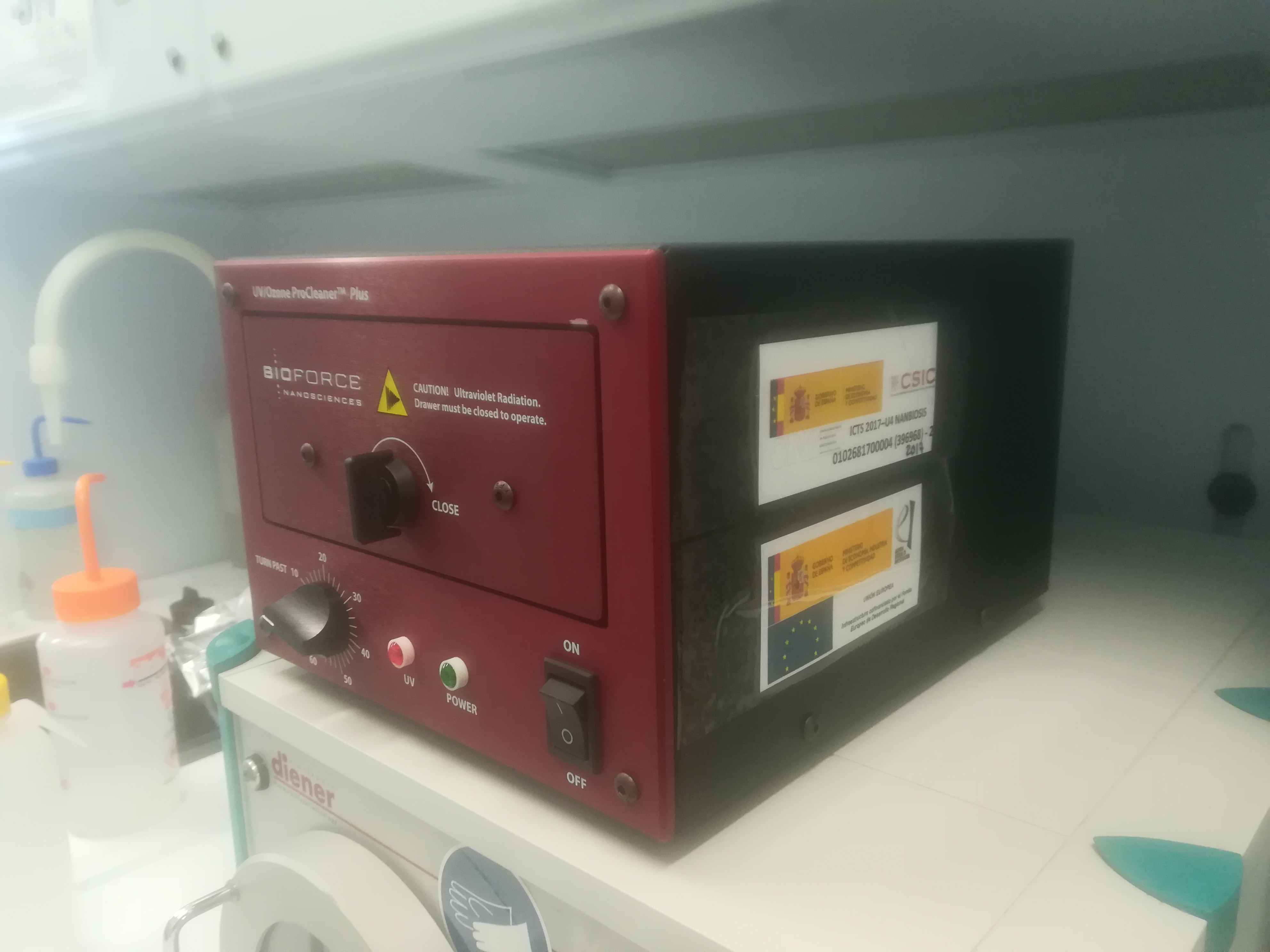
The Biodeposition has been updated with the ozone generation system with a new system (UV / Ozone Cleaner-Procleaner ™), with a lamp with improved power. In the case of the Biodetection unit,  the current device has been to updated in order to make it more competitive. Optical components that improve the performance of the instrument in reference to sensitivity have been changed, the fluidic components has been updated (the flow cell that currently consists of 2 independent channels, peristaltic pumps and injection valves) to increase the automation of the current design and make it more competitive.

European Regional Development Fund

Read More

New laboratory for unit 6 of NANBIOSIS

The equipment of NANBIOSIS U6-Processing of biomaterials and nanoestructuring, that at present are in several laboratories of the ICMAB-CSIC, are going to be transferred to their definitive location in the new laboratory of this NANBIOSIS unit. Given the large amount of equipment the process is expected to last two years, and itwill be done sequentially to continue to provide service to laboratory users. In this process, an engineer has been hired to play a key role, since he will not only be in charge of the transfer, but also to ensure the correct installation of equipment after its transfer and to ensure its start-up and correct operation in the new site.

This action has been confinanced by the European Regional Development Fund (ERDF) through the Plurirregional Operational Program of Spain (POPE)2014-2020 

European Regional Development Fund

Read More

NANBIOSIS Unit 20 expands its equipments and capabilities

NANBIOSIS U20- In Vivo Experimental Platform, led by Dr Simó Schwartz and Dr. Ibane Ibasolo, has recently added new equipment as a result of its participation in the project FICTS1420-20, selected by the MINECO for co-financing by the FEDER Program in ICTS 2014-2020.

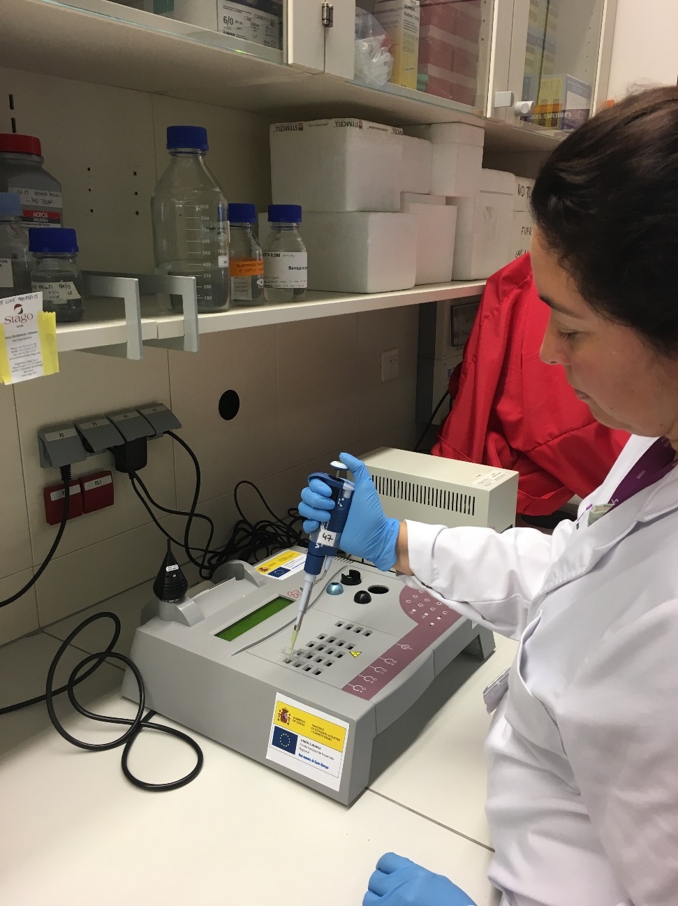
The new equipment is a Coagulometer (Diagnostica Stago STart® 4 Hemostasis Analyzer) used for measuring the  plasma coagulation time upon incubation with different stimulators (PT, APTT, Fibrinogen, D-dimer (quantitative), Thrombin time, Reptilase time etc.) the use o f this equipment will  allow the Unit to test the hemocompatibility of novel nanomedicines in development.

European Regional Development Fund

Read More

Evaluation of a New Design of Antireflux-Biodegradable Ureteral Stent in Animal Model

Francisco Miguel Sánchez Margallo, Assistant Director of NANBIOSIS and Scientific Director of JUMISC, is  co-authors of de article “Evaluation of a New Design of Antireflux-biodegradable Ureteral Stent in Animal Model” published by Urology.

The research were carried out in the NANBIOSIS units in the JUMISC. The purpose was to determine the effects in urinary tract of a new antireflux-biodegradable ureteral stent. Thirty six ureters belonging to 24 pigs were used. The study began with endoscopic, nephrosonographic, and fluoroscopic assessments. Three study groups of ureters (n = 12) were then specified. In group I, a biodegradable antireflux ureteral stent (BDG-ARS) was inserted in the right ureter of 12 pigs. Group II comprised the left ureter of the same animals, in which a double-pigtail stent was placed for 6 weeks. Group III ureters, belonging to 12 additional animals, were subjected to a ureteropelvic junction obstruction model that was then treated by endopyelotomy and stenting with BDG-ARS. Follow-ups were performed at 3-6 weeks and at 5 months.  As result of the research none of the ureters receiving the BDG-ARS showed any evidence of vesicoureteral reflux (VUR). BDG-ARS degradation took place in a controlled and predictable fashion from the third to the sixth weeks, and no obstructive fragments appeared. No differences were found between groups I and II regarding passive ureteral dilation, but significant differences were found regarding VUR and ureteral orifice damage. BDG-ARS always maintained distal ureteral peristalsis. BDG-ARS in group III showed a 50% positive urine culture rate and a 16.6% migration rate in both BDG-ARS groups.

The scientists have demonstrated that morbidity associated with ureteral stents might be reduced as BDG-ARS avoided VUR and bladder trigone irritation, and the polymer combination and stent-braided design achieved a consistent biodegradation rate with no obstructive fragments and with uniform degradation between the third and the sixth weeks. Consequently, morbidity associated with ureteral stents might be reduced.

DOI: 10.1016/j.urology.2018.02.004

Read More