


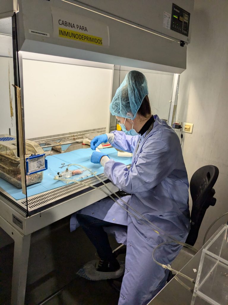
U22-E08. Room to perform small procedures and cures and preparation rooms for large animals prior to the surgical area
Room to perform small procedures and cures and preparation rooms for large animals prior to the surgical area

Room to perform small procedures and cures and preparation rooms for large animals prior to the surgical area
Four multi-use rooms for maintenance of rodents and lagomorphs.
Lagomorphs can individually accommodate upto 102 rabbits.
One barrier zone for rodents maintenance under SPF conditions with capacity for about 3,000 animals in microisolators. It has autoclaves, SAS to the material inlet, automatic beverage system by reverse osmosis, laminar flow hoods for animal handling and integrated operating room within the barrier zone.
One barrier zone for rodents maintenance under SPF conditions with capacity for about 3,000 animals in microisolators. It has autoclaves, SAS to the material inlet, automatic beverage system by reverse osmosis, laminar flow hoods for animal handling and integrated operating room within the barrier zone.
This unit has facilities with a perimeter area of 3,200 m2 and it has large rooms to house the experimental animals according to their species characteristics. Among the equipment and facilities include:
9 rooms for large animals (pigs) maintenance with capacity for about 350 animals, for long periods of time and environmental, nutritional and microbiological conditions controlled.
2 rooms for the maintenance of small ruminants (sheep and goats) with capacity for about 60 animals over long periods of time and environmental, nutritional and microbiological conditions controlled.
JUMISC Animal Housing Unit provides services to companies and research groups in multiple preclinical trials. This unit develops designs and performs preclinical validation on demand. It counts with professionals qualified to design, monitor and validate any type of biomedical study. An important feature of this unit is the possibility to house, maintain and supervise large animals for preclinical validation. This service also counts with a dilated experience in the handling and care of minipigs and what its use in preclinical studies implies.
It has a floor net area of 2.865 m2. It is equipped with spacious rooms to house experimentation animals depending on its specie characteristics (age, sex, weight, breed) and the research projects they are assigned to. Among the equipment and facilities are: rooms for the maintenance of large animals, rooms for the maintenance of small ruminants, rooms for the maintenance of birds, barrier-zone for the maintenance of rodents in SPF conditions, an operating room integrated within the barrier area, multipurpose rooms for the maintenance of rodents and lagomorphs, a room for the reception and handling of the animals, quarantine rooms for pre-experimental processes, a room for small procedures and cures and rooms for the preparation of large animals prior to the surgical area.
Among the services that the Unit develops, we highlight the following:
Customer benefits
All rooms and facilities included in the unit are certified with ISO-9001 and Good Laboratory Practices (GLP), strict quality standards that allow the production of highly accurate results.
In this way, preclinical, security and efficacy studies in animal models can be performed in compliance with the strict guidelines of the regulation agencies, ensuring reliability and traceability of all tests and results performed in its different services.
In addition, this team has been certified by the Spanish Agency of Drugs and Health Products (AEMPS) for studies of “Dosification of problem substances and non-clinical samples”, “Biocompatibility studies of health products”, “In vivo toxicity”, “Tolerance” and “Pharmacodynamics”. All these certifications allow to carry out studies to verify the efficacy, security and biocompatibility of nanotechnological development.
Target customer
The offered services can be of interest to a large number of companies, including the following:
Pharmaceutical and biotechnological companies: Companies that carry out preclinical and clinical studies for the development of drugs, therapies and biotechnological products and require animals to test the efficacy and security of their products.
Academic Researchers: Professors, researchers and students from academic institutions who need access to animals to conduct scientific research in areas like biology, medicine, behaviour, etc.
Medical Institutions: Hospitals, clinics and medical research centres that conduct studies to understand human diseases and develop medical treatments, including preclinical trials on animals.
Medical devices companies: Medical devices manufacturers who need to test the security and efficacy of their products on animals before commercialization.
Regulatory agencies: Government agencies in charge of regulating and monitoring security and efficacy of medical and pharmaceutical products, which may require data from animal studies to support products approval.
Food and agriculture companies: Companies that develop food and agricultural techniques may need to conduct animal studies to evaluate food security, the effects of additives, efficacy in agricultural products, etc.
Additional information
Selected publications:
The aim of this service is to evaluate the in vivo toxicity of nanoparticles and drug-loaded nanoparticles. Thus, we offer different mouse models in which we can determine the toxicity of the compounds. We offer to evaluate the nanotoxicology in healthy mice of any strain or in tumor mouse models. In the service we have subcutaneous and orthotopic/disseminated models of lymphoma, leukemia and colorectal, endometrium, head and neck carcinomas. Moreover, we have also available PDX models of colorectal and endometrium cancer. We can also offer the possibility to set up new animal models of other tumor types.
Customer benefits
The customer will benefit from the experience of the researchers involved in the service that have evaluated the nanotoxicology of a high number of diverse nanoparticles. The service has also high expertise in developing new cancer animal models. Thus, we offer also the possibility of setting up new models of any cancer type that fit with the needs of the customer. Moreover, the conditions of the assays are flexible and will be adapted to the need of each specific compound.
Target customer
The offered service can be of interest to research groups of academia or companies willing to test the nanotoxicology of nanoparticles or drug-loaded nanoparticles in vivo.
References
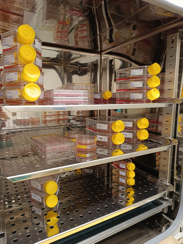
The aim of this service is to evaluate the in vitro toxicity of compounds in different types of normal human cells in which we determine the effect using different types of assays including MTT, LDH release and caspase activation. The cellular models that we offer include cells from kidney, liver, bone marrow stroma or peripheral blood. Additionally, we are offering immunology assays including detection of NO production and chemotaxis and phagocytosis assays.
Customer benefits
The customer will benefit from more than 20 years of experience of the researchers involved in the service that have performed the in vitro evaluation of the toxicity of different nanoparticles and other types of compounds. We offer also the possibility of setting up new in vitro assays or to offer other cellular models that could be of interest for the costumer. Moreover, the conditions of the assays are flexible and will be adapted to the needs of each specific compound.
Target customer
The offered service can be of interest to research groups of academia or companies willing to test the toxicity of any compound in vitro.
References
Leica TCS-SP5 confocal microscope with especial features that allows studying interactions between cells/tissues and materials. Indeed, the experience of the research group in charge of this Unit makes this a unique service for the study of cells and tissues and the interactions between various materials and cell components as well as between implants/scaffolds and tissues of the recipient organism. For example, it makes it possible to characterize in detail the specific markers of particular cell populations for tissue engineering applications and in vitro tests of biocompatibility of new materials, allowing the process of cell colonization of surfaces to be explored and simultaneous analysis of the cells or subcellular structures and materials. The combined use of fluorescence and reflection makes it possible to study the tissue-implant interface, with up to 8 fluorophores, at wavelengths from 405 to 633 nm allowing histological studies of a wide range of tissue types which would be restricted at certain fixed wavelengths due to autofluorescence phenomena.
Leica TCS-SP5 confocal microscope with:
DMI 6000 inverted microscope with 4 objectives:
10X (dry); 20X; 40X and 63X(for immersion).
Confocal module:
•• 3 channels for spectral detection
•• AOBS (Acousto Optical Beam Splitter)
•• Resonant scanning system.
4 lasers:
•• Argon laser (excitation at 458, 477, 488, 496 and 514 nm)
•• He/Ne laser (excitation at 633 nm)
•• DPSS (diode-pumped solid-state) laser (excitation at 561 nm)
•• UV laser (excitation at 405 nm)
Incubation chamber on a motorized stage.
Workstation with 4 types of software for data acquisition and processing:
•• 3D imaging
•• Colocalization analysis
•• FRAP (Fluorescence Recovery After Photobleaching)
•• FLIP (Fluorescene Loss In Photobleaching)
•• FRET (Fluorescence Resonance Energy Transfer)